 EN
EN


Table of Contents
- What Is Canthoplasty? The Three Main Types of Canthoplasty
- Epicanthoplasty: Opening the Inner Corner of the Eye
- Lateral Canthoplasty: Extending the Outer Corner Outward
- Lower Canthoplasty: Softening an Upturned Eye
- Which Type of Canthoplasty Is Right for You?
- How Canthoplasty Is Performed, Step by Step
- Canthoplasty Recovery Time: Week by Week
- Risks, Scarring, and Revision You Should Know About
- Frequently Asked Questions About Canthoplasty
- Planning Your Canthoplasty Consultation in Seoul
Canthoplasty is a group of eye corner procedures that widen the visible opening of the eye by releasing or repositioning the inner corner, the outer corner, or the outer-lower lid line. The three main types of canthoplasty are epicanthoplasty (inner corner), lateral canthoplasty (outer corner), and lower canthoplasty (outer-lower line) — they share a name, but they correct different structures and produce different changes.
I am Dr. Lee Sung-wook, a board-certified plastic surgeon and the director of MINE Plastic Surgery & Dermatology in Seoul. The question I hear most often in my consultation room is simply, “Do I need the inner corner opened, or the outer corner?” This guide walks through what each of the three procedures actually does, which one suits which eye, how recovery unfolds week by week, and the risks I insist every patient understands before deciding.
What Is Canthoplasty? The Three Main Types of Canthoplasty
Canthoplasty refers to surgery on the corners of the eye rather than on the eyelid crease. The surgeon releases a skin fold at the inner corner, or repositions the tissue at the outer corner, so that more of the eye becomes visible. The goal is a wider, more relaxed eye opening that still looks like your own eye.
Why eyes look small or “closed off”
When patients say their eyes look small, the eyeball itself is almost never the issue. What matters is how much of the eye the surrounding structures cover. In my clinical experience, four causes account for most cases:
- An epicanthal fold — a small flap of skin that drapes over the inner corner like a curtain. It is very common in East Asian eyes and hides the natural starting point of the eye.
- Limited horizontal width — the structures at the outer corner limit how far the eye extends sideways, which reads as a crowded, tired expression.
- An upturned outer corner — when the outer corner sits high, the eye can look smaller and the expression can be misread as sharp or severe.
- Ptosis — weakness of the muscle that lifts the upper lid, so the lid covers part of the iris. This is a different problem with a different solution.
There is an anatomical reason this pattern is so common. Anatomy textbooks note that in East Asian eyes the outer corner sits roughly 8 mm higher than the inner corner, and the palpebral fissure (the visible opening between the upper and lower lids) tends to be narrower than it is in Caucasian eyes. That is precisely why lateral and lower corner surgery aims to bring the outer corner down and widen that opening.
Aesthetically, eyes are generally described as balanced when the ratio of eye width : gap between the eyes : eye width is close to 1:1:1. A prominent epicanthal fold hides the start of the eye and breaks that balance — and restoring it is exactly what inner corner surgery is designed to do.
Epicanthoplasty vs lateral canthoplasty vs lower canthoplasty at a glance
| Comparison | Epicanthoplasty | Lateral canthoplasty | Lower canthoplasty |
|---|---|---|---|
| Area corrected | Epicanthal fold at the inner corner | Tissue at the outer corner | Outer-lower lid line |
| Main effect | Horizontal widening, softer gap between the eyes | Horizontal widening, less crowded look | Greater vertical height, softer expression |
| Typically chosen for | Eyes with a clear epicanthal fold and wide spacing | Eyes with enough conjunctival slack at the outer corner | An upturned outer corner that reads as severe |
| Often combined with | Double eyelid surgery | Lower canthoplasty | Lateral canthoplasty |
Epicanthoplasty: Opening the Inner Corner of the Eye
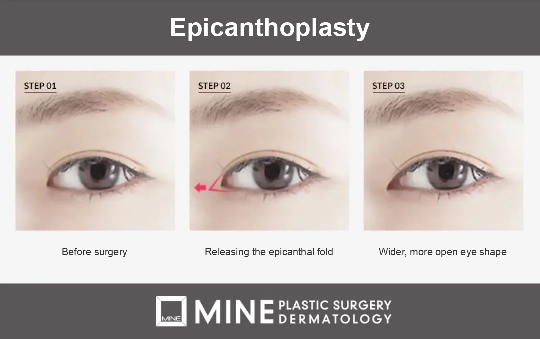
Epicanthoplasty releases and repositions the fold of skin covering the inner corner, so the hidden starting point of the eye is revealed. It widens the eye horizontally at the nose-side end and, at the same time, softens the impression that the eyes sit too far apart. It is the one type of canthoplasty that involves a skin incision, which is why scar planning matters so much.
What actually changes after epicanthoplasty
The measurable gain is modest. Each eye typically widens by about 1–2 mm horizontally. Patients are often surprised that such a small number can change the look of the face, but the perceived change is usually larger than the measurement suggests, because the spacing between the eyes and the front end of the double eyelid line are tidied up at the same time.
Why I design the inner corner conservatively
In my clinical experience, the success of inner corner surgery has little to do with how far you open the corner and everything to do with how naturally the fold is released, without leaving a visible mark. Open it too aggressively and the eyes can look uncomfortably close together, or a strip of pink inner tissue can end up exposed. I design each incision around the patient’s own eye spacing (intercanthal distance) and fold type.
The surgical literature supports that caution. Techniques that redrape the skin can leave an unnatural dimple (a small pressed-in dent) at the point where the fold was released, and older local-flap techniques carry a risk of scar contracture — scar tissue tightening as it heals — which can blunt the angle of the corner. There is no single technique that suits every eye; each carries a trade-off, so the method is chosen to match the anatomy in front of me.
Lateral Canthoplasty: Extending the Outer Corner Outward
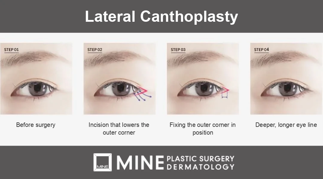
Lateral canthoplasty releases the tissue at the outer corner so that the eye extends further sideways. Because it works behind the lash line rather than through open skin at the front of the eye, scarring is usually less of a concern here — but the amount of widening that is realistically available is far more limited than most patients expect.
How far the outer corner can actually be widened
The outer corner is not a loose flap of tissue. It is held in place by the lateral canthal tendon (the small ligament that anchors the outer corner of your eye to the bone), and the deep part of that ligament attaches firmly to thickened periosteum over Whitnall’s tubercle — a raised ridge on the inner surface of the bone at the side of the eye socket.
In other words, the outer corner is anchored to bone, and that anchoring sets a ceiling on how far it can be extended. How much widening is possible depends on the slack in the conjunctiva (the thin membrane over the white of the eye) at the outer corner and on the shape of the orbital bone. This is why I set expectations during the consultation, before any surgery is booked.
Re-adhesion: why lateral canthoplasty can partly fade
There is one structural property of outer corner surgery that every patient should know. Tissue that has been separated has a natural tendency to reattach during healing, and when that happens the widening effect is partly lost. This is called re-adhesion.
The answer is not to release more tissue; it is to use a fixation and suture design that holds the released tissue in its new position while it heals. When there is not enough conjunctival slack to begin with, pushing the correction further tends to leave redness, a gritty foreign-body sensation, and re-adhesion, without the widening the patient hoped for.
Lower Canthoplasty: Softening an Upturned Eye
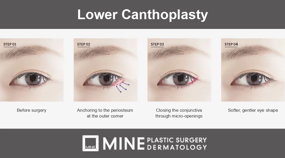
Lower canthoplasty lowers the outer-lower lid line slightly, increasing the vertical height of the eye and softening an upturned, severe-looking outer corner. Of the three types of canthoplasty, this is the one that changes the overall expression most.
Vertical height, not just width
Inner and outer corner surgery both work horizontally. Lowering the outer-lower line works on the other axis, opening the eye vertically at its outer third. It is chosen most often by patients who tell me they look angry when they are not.
Combining lateral and lower canthoplasty (dual canthoplasty)
Because the two work on different axes, combining outer corner and outer-lower corner surgery — often called dual canthoplasty — can widen the eye horizontally and vertically at once, which is why the pair is frequently planned together.
The limiting factor is support. Lower the line too far and the lower lid loses some of its ability to sit snugly against the eyeball, which can lead to exposed inner lid tissue, watering eyes, and — in more pronounced cases — ectropion (the lower lid turning outward). Standard textbooks list the recognized complications of eyelid surgery as scleral show (more white of the eye visible than before), lid retraction, lid laxity (a slack lower lid), and ectropion. These are uncommon, but they are well documented in the literature, so I assess the strength and support of the lower lid first and set the extent of correction from that.
Which Type of Canthoplasty Is Right for You?
Choosing between the three is a diagnostic decision. Here is how the indications separate in practice.
When epicanthoplasty makes sense
Inner corner surgery is a reasonable option when the epicanthal fold is clearly defined, the starting point of the eye is hidden, and the eyes appear widely spaced. It also helps when the front of the double eyelid line rolls inward and looks cramped.
When lateral or lower canthoplasty makes sense
Outer corner surgery is a better fit for eyes with genuine conjunctival slack at the outer end, so there is something to release. Outer-lower correction is the choice for an upturned corner that reads as sharp. When both apply, the two are often planned as one combined procedure.
Male patients: what I plan differently
For male patients I generally plan a more conservative widening. An eye opening that reads as pleasantly large on one face can look artificial on another, and the brow position and orbital shape are usually different as well. If you would like to see how the planning differs, our page on canthoplasty for men covers it in more detail.
When canthoplasty is not the answer
When the eye looks small because the levator muscle is weak, corner surgery is the wrong tool. In that situation ptosis correction is the first-line solution, because it addresses how far the upper lid actually lifts. What many patients do not realize is that the diagnosis has to come first. Once you understand why the eye looks closed off, the choice of procedure usually follows on its own.
How Canthoplasty Is Performed, Step by Step
The sequence below is what a patient at our clinic goes through, from the first measurement to suture removal.
Consultation and measurement
The assessment looks at four things together: how much conjunctival slack there is at the outer corner, the shape of the orbital bone around it, the type and depth of the epicanthal fold, and how strongly the upper lid actually opens. Skipping the last of these is how patients end up with corner surgery when they needed lid elevation.
Design, incision, and fixation
Each of the three procedures follows a different sequence. Inner corner surgery releases the fold and redrapes the skin. In outer corner surgery, the incision brings the corner down, and the tissue is then fixed in the intended position. Outer-lower correction anchors the tissue to the periosteum at the outer corner and then closes the conjunctival portion through micro-openings.
Corner surgery is often planned alongside other eyelid work. If the crease is faint, adding double eyelid surgery gives a more finished result; if the lid is heavy, combining the procedure with ptosis correction usually matters more than widening the corners. You can see how the surgical methods and design process are handled on our canthoplasty procedure page.
How to prepare in the 1–2 weeks before surgery
- Stop smoking and drinking at least one to two weeks beforehand — both directly affect circulation and wound healing.
- Tell us about every medication and supplement you take. Aspirin, omega-3, and similar products can affect bleeding, so bring the list to your consultation and ask your prescribing doctor whether any of them should be stopped.
- Wear glasses instead of contact lenses on the day of surgery, and have eyelash extensions removed in advance.
- Disclose any eye conditions such as dry eye or conjunctivitis, since they change the surgical plan.
Canthoplasty Recovery Time: Week by Week
Canthoplasty recovery time follows a fairly predictable curve. Stitches are usually removed 5–7 days after surgery, and most of the visible swelling settles within one to two weeks. The redness along an incision takes considerably longer to fade than the swelling does.
| Timeframe | What to expect |
|---|---|
| Day 0–3 | Swelling and bruising at their peak; cold compresses recommended |
| Day 5–7 | Stitches removed; gentle face washing from the following day |
| Week 1–2 | Major swelling subsides; most daily activities possible |
| Week 2–4 | Residual swelling and redness decrease; contact lenses usually from this point on |
| Month 3–6 | Scar redness fades and the result settles |
The first two weeks
Use cold compresses for the first two to three days to bring the swelling down, then switch to warm compresses when we tell you to. Keep water away from the surgical site until the stitches are out. Avoid alcohol, smoking, and saunas for at least two to four weeks, as all three slow healing and raise the risk of inflammation.
One habit deserves particular attention: rubbing your eyes. It is a common cause of re-adhesion at the outer corner and of a scar line widening at the inner corner, so it is worth consciously avoiding during the healing period.
When you can wear contact lenses and eye makeup again
Light face washing is usually possible from the day after the stitches come out. Contact lenses and eye makeup are generally resumed about two to four weeks after surgery. Recovery speed varies with the extent of the surgery and with individual healing, so please have your progress checked at the clinic before restarting either.
For at least three months, wear sunglasses and apply sunscreen whenever you go outdoors, so the scar does not darken. If you want the full pre- and post-operative checklist, our guide to eye surgery precautions goes through it in order.
Risks, Scarring, and Revision You Should Know About
Eye corner surgery is performed very often, but it is delicate territory, and you should decide with an accurate picture of what can go wrong. So rather than describe only the upside, I want to set that out honestly.
Scarring and hypertrophic scars
Inner corner surgery involves a skin incision, so a red scar is visible in the early weeks and, depending on your skin type, it can become raised. The redness generally fades over three to six months, and sun protection and scar care during that window strongly influence the final appearance. Some patients need additional help such as scar ointment or laser treatment.
Re-adhesion and over-correction
The three issues I discuss most often are scarring, re-adhesion, and the change in expression that follows over-correction. Over-correction looks different at each corner: too much at the inner corner makes the eyes appear too close together, too much at the outer-lower line can expose inner lid tissue and cause watering or ectropion, and too much at the outer corner leaves redness and a gritty sensation. General surgical risks apply as well — swelling, bruising, inflammation, bleeding, asymmetry, and a temporary worsening of dry eye.
If you are unhappy: reversal and revision
An unsatisfactory result can often be improved through reversal of the inner corner or through eye revision surgery. Reversal is normally assessed only after about six months, once the tissue has stabilized. Revision is more demanding than the first operation because there is less tissue slack to work with, which is exactly why planning the first surgery conservatively is the most reliable form of prevention. No surgeon can promise that both eyes will end up identical; what I can describe honestly is the range of change that is realistic for your anatomy.
Frequently Asked Questions About Canthoplasty
What is the difference between epicanthoplasty and lateral canthoplasty?
The epicanthoplasty vs lateral canthoplasty question comes down to which corner is corrected. Epicanthoplasty works at the inner corner, widening the eye by roughly 1–2 mm per side and tidying the gap between the eyes; lateral canthoplasty widens the eye outward at the outer corner. The inner corner involves a skin incision, so scar care is the priority, while the outer corner is mainly about preventing re-adhesion.
How long is the recovery time after canthoplasty?
Stitches come out at 5–7 days, major swelling settles within one to two weeks, residual swelling and redness continue improving for two to four weeks, and scar redness stabilizes over three to six months. The pace depends on how extensive the surgery was and on individual healing.
Does lateral canthoplasty come undone over time?
Partly true. The separated tissue at the outer corner has a structural tendency to reattach during healing, which can reduce the effect. Accurately assessing conjunctival slack and using a fixation technique that holds the released tissue in position both matter, and avoiding eye rubbing during recovery helps as well.
Will canthoplasty leave a visible scar?
Since an incision is involved, no surgeon can honestly say there will be no scar. The realistic aim for a canthoplasty scar is that the redness fades over three to six months to a point where it is not noticeable unless someone looks closely. Depending on your skin type, scar ointment or laser treatment may be needed to get there.
When can I wear contact lenses and eye makeup again?
Light face washing is usually fine from the day after stitch removal, while contact lenses and eye makeup are generally resumed about two to four weeks after surgery. Because healing varies from person to person, have your progress checked in person before you start again.
Can epicanthoplasty be reversed if I don’t like the result?
Reversal is possible, but it means restoring tissue that has already been incised, so it is more difficult than the original surgery and the outcome is limited by the state of the tissue. It is normally assessed after about six months, once healing has settled — another reason to design the first operation carefully.
Planning Your Canthoplasty Consultation in Seoul
Even for the same procedure, the right combination and the right extent vary considerably from one patient to the next. What decides them is your own anatomy: the shape of your epicanthal fold, the slack in your conjunctiva, the bone structure at the outer corner, and how well your lower lid is supported.
According to the ISAPS Global Survey, eyelid surgery was the most commonly performed cosmetic surgical procedure worldwide in 2024, with about 2.11 million cases. The more routine an operation becomes, the more the outcome depends on the individual surgeon — which makes choosing that surgeon the single most important decision you will make.
How to check that your surgeon is board-certified
In Korea, a board-certified plastic surgeon has completed medical school, a one-year internship, and four years of plastic surgery residency before passing the specialist board examination. You can verify a surgeon’s certification through the Korean Society of Plastic and Reconstructive Surgeons.
Beyond credentials, notice how the consultation is conducted. A surgeon who explains the limits of your anatomy, and how the result will sit with the rest of your face, is giving you the information you need. Cost depends on your eye structure and on which procedures are combined, so we confirm it at your in-person consultation.
If you would like your own eye structure assessed, you can review the surgical options and design approach for canthoplasty at MINE, or call us at +82-2-516-1175 to arrange an in-person consultation in Seoul.
Written and medically reviewed by Dr. Lee Sung-wook, board-certified plastic surgeon, MINE Plastic Surgery & Dermatology.
※ This content is for informational purposes only and does not replace professional medical advice, diagnosis, or treatment. Individual results may vary, and side effects may occur. Please consult with a board-certified plastic surgeon for personalized advice.