EN


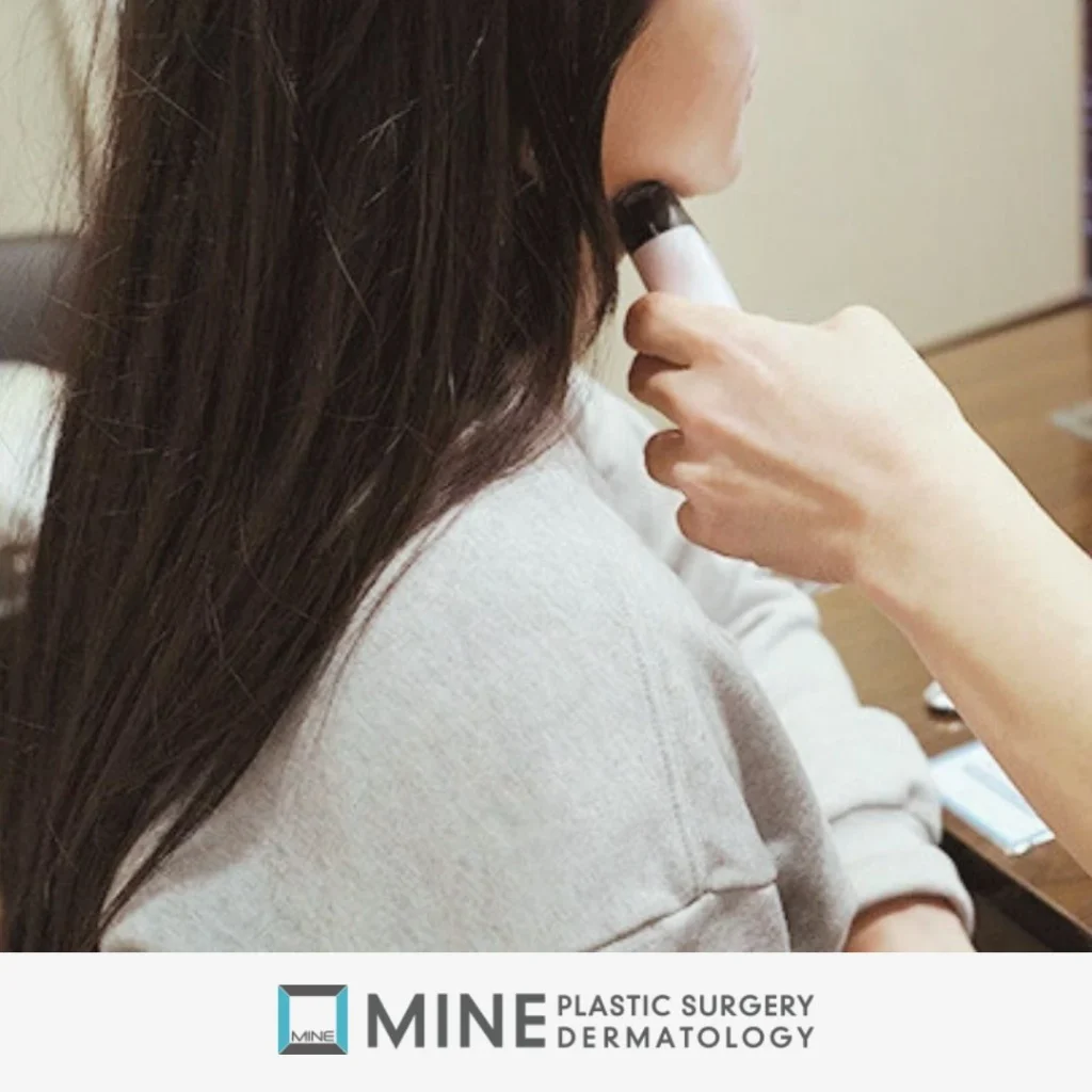
Salivary gland ultrasound is a quick, radiation-free imaging test that uses sound waves to show the size, position and texture of the salivary glands under your jaw. It also tells us whether fullness there comes from the gland, from fat or from the chewing muscle.
Many patients arrive at my clinic convinced they have a stubborn double chin and ready to book liposuction. In my clinical experience, a salivary gland ultrasound quite often shows that an enlarged submandibular gland (the saliva gland tucked under the angle of the jaw) is the main cause. When that is the case, removing fat alone gives only a limited improvement.
This guide focuses on one thing: how the diagnosis is actually made. If you first want to understand what salivary gland hypertrophy is and why it happens, start there and come back.
What Is a Salivary Gland Ultrasound and Why Does It Come First?
A salivary gland ultrasound is the most basic and, in my view, the most important exam for fullness under the jaw. A small handheld probe sends sound waves into the tissue, and the echoes build a live picture on the screen. Nothing is injected, and there is no radiation.
On that live picture we can see the submandibular gland in real time: how large it is, how far it has enlarged, exactly where it sits, which direction it bulges, and where the fat layer ends and the gland begins. Because the scan is short and painless, the treatment direction can usually be discussed at the same visit.
Why the mirror and a pinch test are not enough
From the outside, a soft bulge under the jaw looks much the same whether it is fat, gland or both. A pinch only tells you about the skin and the fat closest to the surface. The gland sits deeper, beneath a thin sheet of neck muscle called the platysma, so you cannot reliably feel its full size with your fingers.
What ultrasound adds to a hands-on exam
A salivary gland ultrasound turns guesswork into a visible map of the layers. Surgical textbooks also point out that ultrasound can tell a solid lump from a fluid-filled cyst and can help spot an abscess (a pocket of pus). CT scanning is kept for specific questions, such as whether a problem starts inside the gland or next to it. MRI adds little when the gland is simply enlarged. For a cosmetic complaint with no warning signs, ultrasound is usually all the imaging you need.
Who Should Have a Submandibular Gland Ultrasound?
You do not need to be sure of the cause before booking a salivary gland ultrasound. Finding the cause is the whole point of the exam.
Signs your fullness may be gland, not fat
- A soft, rounded fullness just under or behind the angle of the jaw, often on both sides
- Fullness that stays put even after weight loss
- A jawline that still looks heavy after previous liposuction under the chin
- You are considering salivary gland Botox or gland reduction and want a scan to guide the decision
The full list of typical signs is covered in our guide to salivary gland hypertrophy. Here we stay with how those signs are confirmed.
When to skip straight to a medical check-up instead
A swelling that is painful, on one side only, hard, or growing quickly is not a cosmetic question. Please read the red-flag section below first. Those symptoms call for an ENT (ear, nose and throat) or medical work-up before any aesthetic treatment is discussed.
What Happens During the Exam: History, Palpation and the Scan
A diagnosis-first consultation follows three simple steps. None of them involve needles.
- History: questions about your habits, health and medications
- Palpation and the clench test: a hands-on check of the jawline and neck
- Salivary gland ultrasound: a live scan of both sides
Step 1: Questions about habits, health and medications
I ask when you first noticed the fullness and whether it has changed. I also ask about diet and alcohol, and whether the area swells or aches around meals. Other questions cover dry mouth or dry eyes, diabetes or other long-term conditions, and any medications you take regularly. These answers help separate a cosmetic gland prominence from a swelling that needs medical attention, and they tell me what to look for on the salivary gland ultrasound.
Step 2: Palpation and the clench test
Next, I feel the area under the jaw and watch how the jawline and neckline move as you swallow and turn your head. Then comes a simple trick I use at almost every consultation: the clench test.
Bite down firmly on your back teeth. If the area becomes hard and pushes out, the masseter (the main chewing muscle) is the likely cause. If it stays soft, the salivary gland is the more likely suspect. Medical literature describes the visual difference the same way: a large masseter makes the face look square and angular, while a large gland makes the lower face look round and heavy.
Step 3: The ultrasound scan itself
You lie back with your chin slightly raised. A little gel goes on the skin, and the probe glides along the area under the jaw. Both sides are scanned and compared. The scan is painless, uses no radiation, and you can watch the screen while I explain what each layer is.
What a Salivary Gland Ultrasound Shows: 5 Key Findings
This is the heart of the diagnosis. A salivary gland ultrasound is read layer by layer. The surgical literature on submandibular sialadenosis and dedicated ultrasound textbooks describe what to look for. Below are the five findings I check, in plain language.
1. The layers: skin, fat, fascia, gland and muscle
An ultrasound textbook describes 5 layers on a normal scan of this area: skin, fat, fascia (a thin connective-tissue wrapping), gland tissue and the mylohyoid muscle (the floor-of-mouth muscle beneath the gland). The submandibular gland normally looks a little darker on screen than the parotid gland in front of the ear. Doctors call this “more hypoechoic,” which simply means it reflects fewer echoes. The facial artery runs behind or through the gland, so we also note where it lies.
2. Gland bulk and which way it protrudes
On a salivary gland ultrasound, size is judged in context, not against a single number. I compare the left and right sides, check how far the gland drops below the jaw line, and weigh its bulk against the fat lying over it. Just as important is the direction of the bulge. A gland pushing outward and downward changes the jawline far more than one sitting deep and high.
3. Uniform texture with no lump
When a gland is enlarged for non-inflammatory reasons (a condition called sialadenosis), the ultrasound textbook describes a bigger gland with an even, uniform texture and no separate lump inside it. That uniform look is reassuring. In a painless swelling on both sides, it argues against a tumor or a blocked duct.
4. Blood-flow (Doppler) signal
Doppler mode, a setting on the same ultrasound machine, shows blood flow as color on the screen. Normal or low flow in the gland argues against active inflammation. A gland with heavy blood flow, especially with pain or fever, points toward infection rather than a cosmetic issue.
5. Where the fat sits relative to the platysma and gland
Fat can lie above the platysma muscle, below it, or both, and the gland sits deeper still. On a salivary gland ultrasound, each layer shows up separately, so we can see whether fat, gland or a mix of both is behind the fullness. For the broader comparison, see double chin vs salivary gland. If the scan shows mostly fat, our article on ultrasound consultation for double chin surgery explains that side of the picture.
| Ultrasound finding | What it suggests | Typical next step |
|---|---|---|
| Enlarged gland, even texture, no lump, normal blood flow | Cosmetic gland prominence or sialadenosis | Discuss Botox or gland reduction |
| Thick fat layer, gland of ordinary bulk | Fullness mainly from fat | Fat-focused treatment |
| Both a prominent gland and a thick fat layer | Mixed cause | Combined plan |
| Separate lump, bright spot with a shadow, or strong blood flow | Possible tumor, stone or inflammation | ENT or medical work-up first |
How Is Salivary Gland Hypertrophy Diagnosed vs Other Causes of Swelling?
Salivary gland hypertrophy is diagnosed when the gland is enlarged with an even texture, no lump and no signs of infection, and when medical causes have been ruled out. Most people who come to us for a heavy jawline have a cosmetic gland prominence, not a disease. Still, ruling out a disease is always the first job of a salivary gland ultrasound.
Red flags that need an ENT or medical work-up first
- A lump on one side only, especially if it feels hard or fixed
- A swelling that grows quickly over weeks
- Pain or swelling that spikes at mealtimes (a possible salivary stone)
- Fever, redness, tenderness or pus in the mouth (possible infection)
- Dry eyes together with a dry mouth (possible Sjögren’s disease, an autoimmune condition)
- A new swelling that started after a new medication
- Weakness or numbness of the face
Conditions ultrasound helps rule out
A clinical textbook on salivary gland disorders reports that ultrasound detects salivary stones with about 90% sensitivity and 95% specificity. On the screen, a stone appears as a bright spot that casts a dark shadow behind it, although very small stones can be missed. Surgical textbooks list other causes of gland swelling to exclude. These include bacterial or viral infection (such as mumps), granulomatous conditions (such as tuberculosis or sarcoidosis), autoimmune disease (such as Sjögren’s or lupus), metabolic or hormonal disorders, certain drugs, and tumors, most often benign ones such as a pleomorphic adenoma.
According to a 2024 surgical textbook, about 50% of sialadenosis cases are linked to a recognized risk factor such as diabetes, metabolic syndrome, heavy alcohol use, an eating disorder, malnutrition or liver disease. That is why the history in Step 1 matters. If an eating disorder is involved, it is treated first. Surgery is reserved for a significant cosmetic concern.
When CT, MRI or blood tests are added
Textbook descriptions note that in sialadenosis the secretory (acinar) cells, the cells that make saliva, swell to roughly twice their normal diameter. That cell-level change is why the whole gland enlarges evenly rather than forming a lump. Because a salivary gland ultrasound and the clinical picture usually make the diagnosis, a biopsy is rarely needed. CT, MRI or blood tests are added only when something does not fit, such as a lump, a suspected abscess, or signs of an autoimmune or systemic condition.
From Diagnosis to Treatment: What Your Ultrasound Results Mean
Your salivary gland ultrasound result points toward one of a few paths. Our guide to salivary gland hypertrophy treatment options covers each one in depth. Here is the short version.
Mild, soft gland bulk
A modestly enlarged gland is often a candidate for Botox injections first. How that compares with surgery is explained in salivary gland Botox vs surgery.
A large or drooping gland
When the scan shows a large gland bulging well below the jaw line, injections have clear limits. Partial gland reduction is usually discussed instead; see salivary gland reduction surgery.
Gland plus fat or loose neck skin
If the scan shows a prominent gland together with a thick fat layer or loose skin, the plan may combine gland treatment with liposuction or a neck lift. For what comes after treatment, see our articles on recovery time after salivary gland surgery, scarring and cost in Korea.
Frequently Asked Questions About Salivary Gland Ultrasound
How is salivary gland hypertrophy diagnosed?
It is diagnosed by combining a health history, a hands-on exam with the clench test, and a salivary gland ultrasound. The scan should show an enlarged gland with an even texture and no lump. Medical causes such as infection, stones, autoimmune disease or a tumor are ruled out first, so the diagnosis rests on the full picture rather than on size alone.
Can an ultrasound tell a double chin from an enlarged salivary gland?
Yes. Ultrasound shows the fat layer and the gland as separate layers, so it reveals whether fullness comes from fat, gland or both. That distinction matters because liposuction alone gives only a limited result when the gland is the main cause. For the broader differences, see our double chin vs salivary gland guide.
Does a salivary gland ultrasound hurt or use radiation?
No. A salivary gland ultrasound uses sound waves, not X-rays, so there is no radiation. A little gel goes on the skin and a smooth probe glides under the jaw. There are no needles, the scan is short, and the findings can usually be discussed at the same visit.
What does a normal submandibular gland look like on ultrasound?
An ultrasound textbook describes 5 layers on a normal scan: skin, fat, fascia, gland tissue and the mylohyoid muscle. The submandibular gland usually looks slightly darker than the parotid gland and has an even texture with no separate lump. Size is judged by comparing both sides and the surrounding layers rather than by a single number.
Can ultrasound find salivary gland stones?
Usually, yes. A clinical textbook reports that ultrasound detects salivary stones with about 90% sensitivity and 95% specificity. Stones show up as bright spots with a shadow behind them, but very small stones can be missed. If your swelling flares at mealtimes, please see an ENT specialist.
When do I need a CT or MRI instead of an ultrasound?
CT or MRI is added when ultrasound or the exam raises a specific question, such as a lump, a suspected abscess, or disease near the gland rather than in it. Surgical textbooks note that CT helps tell problems inside the gland from those next to it, while MRI adds little for a simply enlarged gland.
When should a swollen salivary gland be checked by an ENT first?
See an ENT or your doctor first if the swelling is on one side and hard, grows quickly, hurts at meals, or comes with fever or redness. The same applies to dry eyes with a dry mouth, or to facial weakness or numbness. These signs need a medical work-up before any cosmetic treatment.
Book a Salivary Gland Ultrasound Consultation at MINE
At MINE Plastic Surgery in Seoul, Dr. Lee Sung-wook, a board-certified plastic surgeon, performs the salivary gland ultrasound and the hands-on exam in the same consultation. You see the layers on screen, hear what they mean, and leave with a clear answer about whether your fullness is gland, fat or muscle, before any treatment is planned.
To learn more about the procedure itself, visit our salivary gland reduction at MINE page. To book a consultation, call +82-2-516-1175 or reach our international patient team through WhatsApp or the online consultation form.
※ This content is for informational purposes only and does not replace professional medical advice, diagnosis, or treatment. Individual results may vary, and side effects may occur. Please consult with a board-certified plastic surgeon for personalized advice.