EN
EN

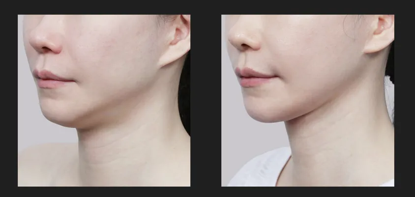
Table of contents
Submandibular gland removal is the surgical removal of one or both submandibular salivary glands, the pair of glands sitting under your jaw that make most of your resting saliva. The biggest worry for most patients is simple: will my mouth and jaw still work normally afterward? Common concerns include lasting dry mouth, less saliva, and nerve injury that affects the lip or tongue.
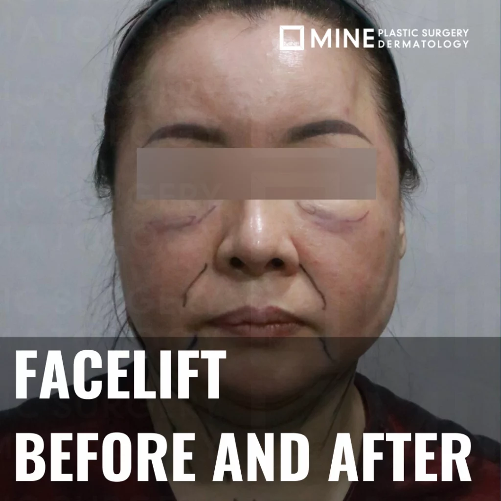
The short answer is reassuring. Because saliva comes from several glands working together, the body compensates well when one gland is taken out, so most functional changes are temporary. Below we explain what really happens to saliva, sensation, and the lip-moving nerves after submandibular gland reduction, based on clinical evidence and modern surgical technique. For day-by-day healing, see our complete recovery guide, and for visual outcomes see these before-and-after results.
What is Submandibular Salivary Gland Removal Surgery?
Submandibular salivary gland removal (also called submandibular gland excision) is the partial or complete surgical removal of the submandibular salivary glands beneath the jaw. Surgeons do this for two broad reasons. The most common medical reason is a blocked or repeatedly infected gland, usually from a salivary stone (sialolithiasis) that cannot be cleared any other way. The cosmetic reason is a heavy or bulging gland that softens the jawline and creates fullness under the chin, often reduced during a double chin contouring procedure.
An important distinction shapes your risk: complete removal takes the whole gland, while partial (subtotal) removal leaves a healthy rim of gland behind. For cosmetic cases, surgeons usually choose partial removal. In published surgical experience, subtotal removal has produced very few of the classic complications, no nerve injury, no fluid collection, and no salivary leak, because the safest tissue is left untouched. That is the main reason functional problems are uncommon when an experienced surgeon performs the procedure.
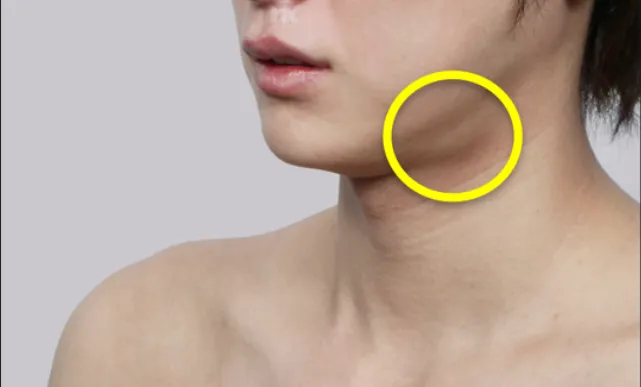
Cosmetic Applications
Aesthetic purposes may include:
- Jawline contouring for facial symmetry
- Neck profile enhancement during facelifts
- V-line facial sculpting procedures
- Double chin reduction as a complementary treatment
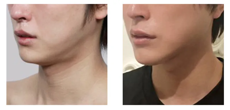
When one salivary gland is removed, the others can provide adequate saliva to help you talk, eat, and swallow. Dry mouth after submandibular gland excision is very rare. This reassuring finding from Johns Hopkins Medicine highlights the body’s remarkable ability to compensate for the loss of one salivary gland.
Expected Functional Changes After Surgery
Immediate Post-Surgical Changes
Following submandibular salivary gland removal, patients typically experience these temporary changes:
- Swelling and bruising around the surgical site (1-2 weeks)
- Mild sensation changes in the jaw area
- Temporary dry mouth during initial recovery days
- Minor swallowing discomfort due to local swelling
Compensatory Mechanisms
The human salivary system demonstrates remarkable adaptive capacity:
- Parotid glands (ear region): Increase saliva production to compensate
- Sublingual glands (under tongue): Maintain or slightly increase function
- Minor salivary glands: Over 500 small glands throughout the mouth contribute to overall saliva production
- Remaining submandibular salivary gland: If only one side is removed, the opposite gland often increases output
Clinical studies show that if one gland is surgically removed, you will not likely notice any degree of dry mouth because the remaining salivary glands compensate effectively.
Impact on Saliva Production
Normal Salivary Function
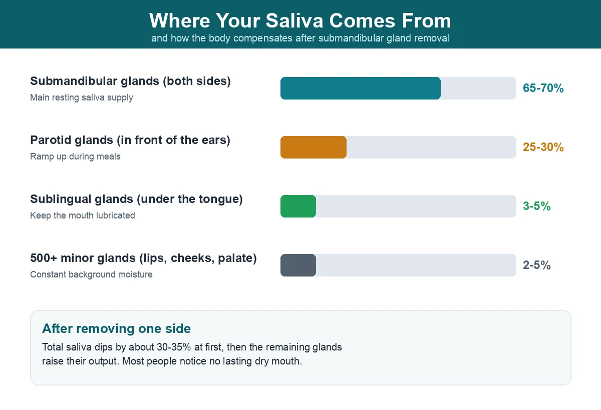
Healthy adults produce approximately 1-1.5 liters of saliva daily. The contribution from each gland type:
| Salivary Gland | Production Percentage | Primary Function |
|---|---|---|
| Submandibular (both sides) | 65-70% | Baseline saliva production |
| Parotid glands | 25-30% | Increased output during meals |
| Sublingual glands | 3-5% | Oral lubrication |
| Minor salivary glands | 2-5% | Continuous moisture |
Post-Surgical Saliva Changes
Unilateral (one-sided) removal:
- Overall saliva reduction: 30-35%
- Compensatory increase from remaining glands
- Most patients experience no significant dry mouth
Remarkable outcomes with partial removal:
Recent clinical research reveals exceptionally promising results for partial submandibular salivary gland removal. Submandibular salivary gland reduction was performed in 112 of 736 consecutive face lifts between 2002 and 2013, with studies showing that partial removal techniques result in zero cases of permanent dry mouth among patients.
This outstanding outcome occurs because:
- Remaining gland tissue compensates through increased function
- Other salivary glands become more active
- Minor salivary glands throughout the mouth enhance production
- Neural regulation remains intact, maintaining normal saliva control
Managing Temporary Dry Mouth
If mild dry mouth occurs initially, these strategies help:
Immediate relief methods:
- Adequate hydration (2+ liters daily)
- Sugar-free gum to stimulate saliva flow
- Artificial saliva products as needed
- Room humidifiers to maintain moisture levels
Long-term management:
- Regular dental checkups for oral health monitoring
- Fluoride toothpaste to prevent decay
- Avoid irritating foods (spicy, acidic, or very salty)
- Excellent oral hygiene practices
Temporary Side Effects and Management
Common Side Effects and Frequencies
Mild complications (10-20% of patients):
- Surgical site swelling and bruising
- Temporary pain and discomfort
- Minor sensation changes around the jaw
Moderate complications (5-10% of patients):
- Hematoma formation (blood collection)
- Seroma development (fluid accumulation)
- Mild temporary dry mouth
Rare complications (1-5% of patients):
- Infection at the surgical site
- Salivary fistula (abnormal connection)
- Excessive scar tissue formation
Effective Side Effect Management
Swelling control:
- Cold compresses for first 48 hours
- Elevated head position during rest
- Anti-inflammatory medications as prescribed
Pain management:
- Prescribed pain relievers taken regularly
- Soft diet during initial recovery
- Gentle jaw movements to prevent stiffness
Infection prevention:
- Antibiotic compliance as directed
- Surgical site cleanliness
- Complete smoking/alcohol cessation
Nerve Damage Risk and Prevention
Vulnerable Nerves During Surgery
Marginal mandibular nerve (facial nerve branch):
- Risk: Temporary mouth corner drooping
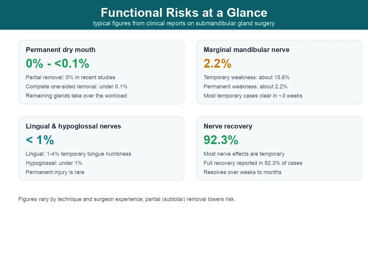
- Frequency: temporary neuropraxia with full recovery of the marginal mandibular nerve following submandibular salivary gland excision occurred in 15.6% of cases, while permanent marginal weakness occurred in 2.2% of cases
- Recovery: temporary cases showed complete spontaneous recovery within about 3 weeks
Lingual nerve:
- Function: Controls tongue sensation
- Risk frequency: 1-4% temporary numbness
- Recovery period: Several months to one year
Hypoglossal nerve:
- Function: Controls tongue movement
- Risk frequency: Less than 1%
- Severity: Rare but potentially more serious if damaged
In plain terms, the nerve that matters most for appearance is the marginal mandibular nerve, which lifts the corner of the lower lip. It runs close to the top edge of the gland, so a careful surgeon works just below it and keeps it in view throughout. The lingual and hypoglossal nerves sit deeper and are protected by leaving a healthy layer of tissue in place, one more advantage of partial removal.
Advanced Nerve Preservation Techniques
Modern surgical approaches have dramatically reduced nerve injury risks:
Facial nerve monitoring:
- Facial nerve monitoring techniques during the procedure protect the marginal mandibular nerve, which controls the muscles of the chin and lower lip
- Real-time nerve identification during surgery
- Electrical stimulation testing to confirm nerve location
Anatomically-guided approaches:
- Subplatysmal dissection planes that avoid nerve pathways
- Careful tissue layer separation maintaining natural barriers
- Microsurgical techniques for precise gland removal
Surgeon experience factor:
The skill and experience of your surgeon significantly impacts outcomes. Use of the nerve stimulator (Parson’s McCabe) or Checkpoint nerve stimulator facilitates identification of the marginal mandibular nerve through stimulation, representing the advanced techniques used by experienced practitioners.
Possibility of Permanent Problems
Realistic Risk Assessment
Permanent dry mouth:
- Partial removal: 0% based on recent studies
- Complete unilateral removal: Less than 0.1%
- Bilateral removal: Higher risk, requires careful consideration
Permanent nerve damage rates:
- Marginal mandibular nerve: permanent marginal weakness occurred in 2.2% of cases
- Lingual nerve: Less than 0.5%
- Hypoglossal nerve: Less than 0.1%
These low complication rates reflect modern surgical techniques and proper patient selection.
Minimizing Permanent Complications
Pre-surgical optimization:
- Comprehensive medical evaluation including imaging studies
- CT or MRI imaging to map anatomical variations
- Patient health optimization (diabetes control, smoking cessation)
Surgical technique excellence:
- Nerve preservation protocols consistently followed
- Adequate surgical exposure for safe gland removal
- Avoiding excessive tissue manipulation
Post-surgical care:
- Regular follow-up monitoring for early problem detection
- Prompt intervention if complications arise
- Rehabilitation therapy when indicated
Recovery Process and Functional Restoration
Step-by-Step Recovery Timeline
Immediate post-surgery (1-3 days):
- Peak swelling and discomfort
- Mild dry mouth possible (especially with complete removal)
- Soft diet recommended
Early recovery (1-2 weeks):
- Progressive swelling reduction
- Gradual activity resumption
- Saliva production normalization begins
Mid-term recovery (2-8 weeks):
- Functional recovery nearly complete
- Compensatory mechanisms fully activated
- Normal eating typically resumed
Complete recovery (3-6 months):
- All functions stabilized
- Scarring minimized
- Final surgical results apparent
Accelerating Functional Recovery
Active oral care:
- Saliva stimulation exercises (sugar-free gum, sour candies)
- Gentle oral massage to promote circulation
- Consistent hydration habits
Nutritional support:
- High-protein foods for tissue healing
- Vitamin C and E supplementation for wound recovery
- Omega-3 fatty acids to reduce inflammation
Frequently Asked Questions
Q: Will I have permanent dry mouth after submandibular salivary gland removal?
A: No, especially with partial removal techniques. Recent clinical studies show 0% permanent dry mouth with partial submandibular salivary gland removal. Even with complete unilateral removal, permanent dry mouth occurs in less than 0.1% of cases due to effective compensation by remaining salivary glands.
Q: When can I eat normally after surgery?
A: Most patients resume normal eating within 1-2 weeks. Initially, soft foods are recommended, but as swelling subsides, regular diet becomes comfortable. Complete functional recovery typically occurs within 4-8 weeks.
Q: Will removing one gland affect my other salivary glands?
A: No, surgery doesn’t directly harm other glands. In fact, remaining salivary glands become more active to compensate for the removed tissue, maintaining overall saliva production effectively.
Q: Is nerve damage recovery possible after submandibular salivary gland surgery?
A: Yes, most nerve injuries recover completely. Neurapraxia of the mandibular branch of the facial nerve was common after surgery, but function was restored in 92.3% of cases. Even when temporary nerve effects occur, the vast majority resolve within weeks to months.
Q: How visible will the surgical scar be?
A: Scars are minimal and well-hidden. The incision is typically 2 to 3 centimeters long and sits under your jaw, where it is unlikely to be noticeable. With proper care, scars fade significantly within 3-6 months and become barely noticeable.
Q: Who is a good candidate for submandibular gland removal?
A: Good candidates fall into two groups. Medically, people with a gland that is repeatedly blocked or infected, usually by a stone, often benefit from removal. Cosmetically, healthy adults with a visibly full or bulging gland that blurs the jawline are typical candidates for partial submandibular gland removal. A consultation with imaging confirms whether the gland is truly the cause of lower-face fullness before any surgery is planned.
Q: Does submandibular gland removal change my voice or taste?
A: No, voice and taste are not affected by the surgery itself. The submandibular gland does not control the vocal cords, and taste is carried by separate nerves. The lingual nerve near the gland can cause temporary tongue numbness in a small number of cases, but true taste loss is not a recognized outcome of submandibular gland removal. Most patients eat, speak, and taste normally once early swelling settles.
Conclusion: Safe and Successful Submandibular Gland Removal
Submandibular gland removal surgery has an excellent safety profile with minimal risk of serious functional problems. The combination of advanced surgical techniques, natural compensatory mechanisms, and expert post-operative care ensures successful outcomes for the vast majority of patients.
Key takeaways for optimal results:
- Modern partial removal techniques eliminate permanent dry mouth risk
- Advanced nerve preservation methods minimize complications
- Skilled surgical technique is crucial for safe outcomes
- Proper post-operative care ensures smooth recovery
- Patient cooperation accelerates healing and recovery
If you’re considering submandibular salivary gland surgery, thorough consultation with an experienced surgeon is essential for proper evaluation and treatment planning. At mineclinic, our specialists utilize the latest surgical techniques and provide comprehensive pre- and post-operative care to ensure optimal outcomes.
For professional consultation about submandibular gland removal and to explore your treatment options, contact mineclinic today.
This information is provided for educational purposes and does not replace professional medical advice. Individual results and risks vary. Always consult a qualified healthcare provider for accurate diagnosis and personalized treatment recommendations.
External References:
- American Society of Plastic Surgeons – Professional surgical guidelines and patient resources
- Johns Hopkins Medicine – Submandibular gland excision clinical protocols
- Mayo Clinic – Salivary gland disorder treatment guidelines
※ This content is for informational purposes only and does not replace professional medical advice, diagnosis, or treatment. Individual results may vary, and side effects may occur. Please consult with a board-certified plastic surgeon for personalized advice.