EN
EN

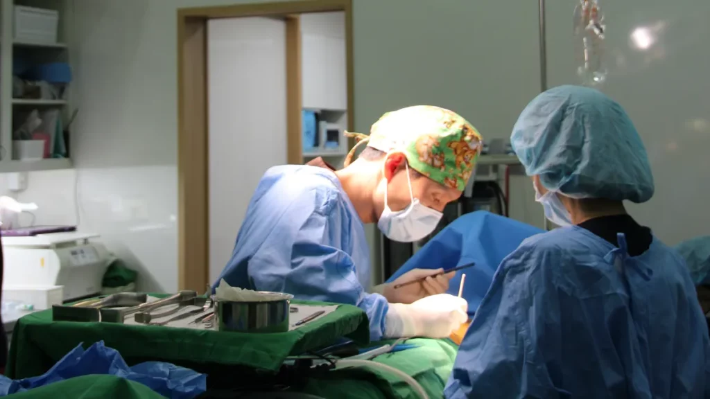
Endoscopic breast augmentation benefits come from one change: the surgeon creates the implant pocket while watching it on a screen, instead of working partly by feel. That direct view makes bleeding control and pocket shaping more precise. It does not, however, make every risk disappear.
I am Dr. Lee Sung-wook, a board-certified plastic surgeon and the director of Mine Plastic Surgery in Gangnam, Seoul. Two questions come up in almost every breast consultation: “Isn’t every endoscope the same?” and “Is it actually safer?” Both are fair questions, and the honest answer has two parts.
In this article I go through the five benefits one by one. For each, I explain which surgical step the camera actually changes, whether the benefit is supported by plastic surgery textbooks or is our own clinical explanation, and what it does not promise. Then I cover the limits, because a camera cannot fix every problem.
- What Endoscopic Breast Augmentation Benefits Actually Mean
- Why Pocket Precision Decides the Result
- HD vs Standard Endoscope: Mine’s Own Comparison
- The 5 Endoscopic Breast Augmentation Benefits, and What Each Does Not Promise
- Limits of Endoscopic Breast Augmentation: What a Camera Cannot Fix
- Recovery Timeline and Preparation
- Frequently Asked Questions
- Planning Your Consultation
What Endoscopic Breast Augmentation Benefits Actually Mean
An endoscope is a thin camera with its own light source. In endoscopic breast augmentation, it is passed through the same small incision used for the surgery, usually in the armpit. The image appears on a monitor, so the surgeon can see the inside of the pocket (the space where the implant will sit) while creating it.
Three terms will come up often in this article, so here they are in plain language:
- Pocket: the space under the breast tissue or chest muscle that holds the implant.
- Dissection: the surgical work of separating tissue layers to create that pocket.
- Hemostasis: stopping bleeding, usually by sealing small vessels with a heated instrument.
Blind dissection vs direct vision: the one difference that matters
When breast surgery is performed through a small incision far from the breast, part of the pocket lies out of the surgeon’s direct line of sight. Without a camera, that part is created by touch and experience. Plastic surgery textbooks note that when a pocket is made bluntly from a distant access point, controlling bleeding can be difficult.
The endoscope changes this. The standard reference textbook Neligan’s Plastic Surgery (Breast volume) describes the endoscope as allowing precise release of the lower attachments of the pectoralis major (the large chest muscle), along with direct visualization for bleeding control, while leaving no scar on the breast itself. For readers who want to go further, the Archives of Plastic Surgery has published a review of transaxillary endoscopic breast augmentation.
Where the camera goes and what the surgeon sees
Through the armpit incision, a short tunnel is made toward the chest. The surgeon first identifies the correct tissue layer, then slides the endoscope through the tunnel into that layer. From there, the dissection continues on screen using a long, insulated electrocautery instrument (a tool that cuts and seals vessels at the same time).
On the monitor, the surgeon sees the muscle fibers, the pocket walls and any small vessels before they are touched. In other words, the camera does not add a new operation. It lets the surgeon see the steps that used to be done partly blind.
Why Pocket Precision Decides the Result
In my clinical experience, most problems after breast augmentation trace back to one stage: how precisely the pocket was made. Each common complication links to a specific mistake during dissection.
- Unseen vessel → bleeding during or after surgery, which can collect as a hematoma (a pool of blood around the implant).
- Unequal left and right pockets → asymmetry in height or shape.
- Wrong pocket boundary or tissue layer → implant malposition or rotation.
- Rough or bloody pocket surface → in our clinical explanation, a higher risk of capsular contracture.
Bleeding and hematoma
Plastic surgery textbooks advise against blind or blunt pocket dissection without proper bleeding control, because it raises the risk of hematoma. This is where the camera helps most directly: a vessel that can be seen can be sealed before it bleeds.
Asymmetry and implant malposition
Textbooks stress that the surgeon must confirm the correct plane before going further. The plane is either under the fascia (the thin film covering the muscle) or under the muscle itself. Textbooks also note that staying in the shallow layer until the outer edge of the chest muscle protects the intercostobrachial nerve, which supplies sensation to the inner upper arm.
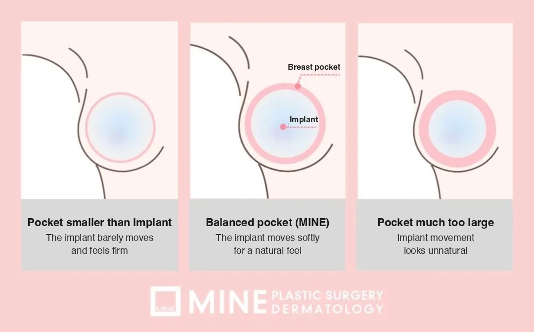
When the boundary of the pocket is misjudged, the implant can sit too high, drift to the side or rotate. If you want to understand this problem in more detail, I have written a separate article on breast implant malposition.
Capsular contracture: what is and is not proven
Capsular contracture happens when the natural scar capsule around an implant thickens and tightens, making the breast firm or changing its shape. Many factors are involved. Our clinical explanation is that a cleaner, less bloody pocket is likely to lower the risk. However, I cannot point to a textbook figure showing that endoscopy reduces capsular contracture by a specific amount, so I will not give you one. For a fuller explanation, see capsular contracture symptoms and prevention.
Dr. Lee’s key point: The result depends less on how large the implant is and more on how precisely the space was made.
HD vs Standard Endoscope: Mine’s Own Comparison
Patients often ask whether an “HD endoscope” is a meaningful difference or just a label. The table below is our own comparison of the system we use with a standard-definition endoscope. Please read it as a description of equipment, not as the result of a published study.
What “HD” changes on the screen
| Feature | Standard endoscope | HD endoscope (Mine’s system) |
|---|---|---|
| Resolution | Standard definition (SD) | Full HD to 4K |
| Viewing angle | Limited | Wide angle, adjustable direction |
| Light source | Standard LED | High-output cold LED light |
| Small-vessel visibility | Harder to identify | Designed to make small vessels easier to identify |
| Pocket-surface check | Partial | Whole dissection viewed in real time |
Based on Mine’s equipment specifications; not a published head-to-head study.
A sharper, brighter image does not operate on its own. What it changes is how easily the surgeon can spot a small vessel or an uneven area on the pocket wall before moving on.
How Mine uses the HD system in five steps
At our clinic, the HD endoscope is part of the standard protocol for breast augmentation through the armpit. The workflow has five stages:
- Planning with a 3D body scan: before surgery, a 3D scan of the chest is used to plan the pocket.
- Real-time monitoring: the whole dissection is performed while watching the monitor.
- Vessel-level bleeding control: small vessels are sealed as they are identified.
- Left-right balance check: both pockets are compared and adjusted for depth and width.
- Final check before closing: implant position, rotation and any remaining bleeding are checked once more.
The last stage matches what textbooks describe as standard practice. A typical textbook sequence includes two safety steps just before the implant goes in: a final check for bleeding and rinsing the pocket with an antibiotic solution.
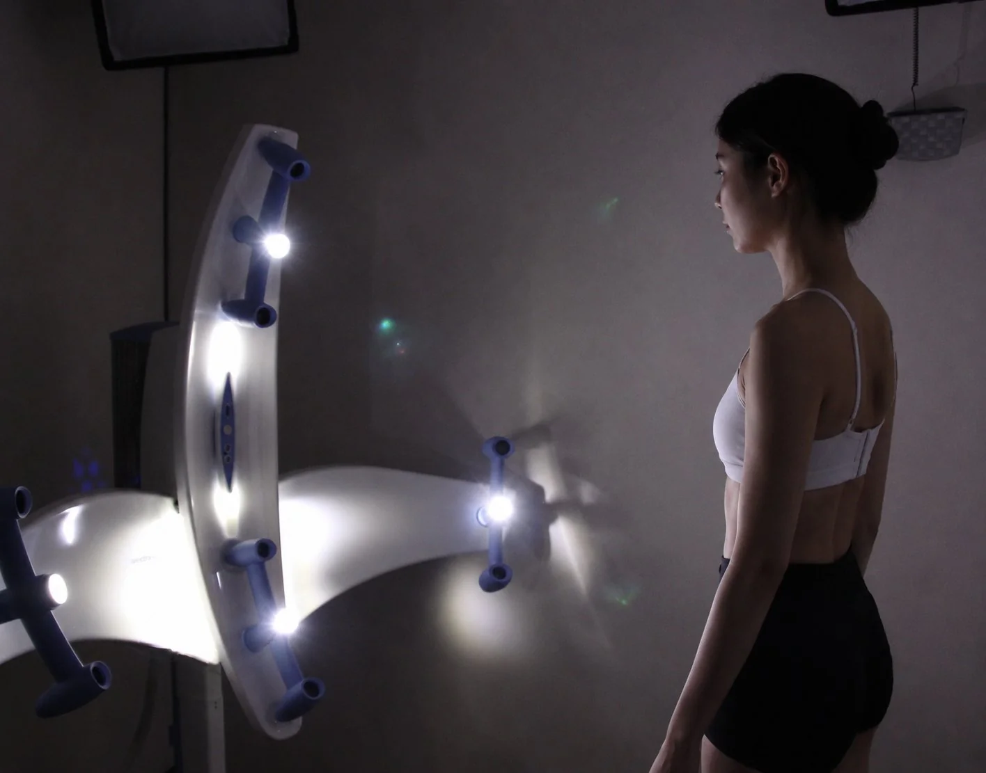
We call a pocket finished this way a “dry pocket,” meaning one with no oozing blood left behind. I explain that concept and its steps on our HD endoscopic breast augmentation at Mine page, so I will not repeat it here.
The 5 Endoscopic Breast Augmentation Benefits, and What Each Does Not Promise
Here are the five endoscopic breast augmentation benefits I discuss with patients. For each one, I separate what textbooks support from what is our own clinical explanation.
1. Lower risk of bleeding and hematoma
What the camera changes: vessels are seen and sealed during dissection, instead of being found only after they bleed.
Evidence level: textbook-supported. Direct-vision hemostasis is one of the main advantages textbooks list for the endoscopic approach, and avoiding blind dissection is recommended to limit hematoma.
What it does not promise: zero hematoma. Bleeding can still occur after surgery, for example when blood pressure rises or a sealed vessel reopens.
2. A cleaner, more even pocket
What the camera changes: the lower attachments of the chest muscle are released in a controlled way while the surgeon watches, and the left and right pockets can be compared on screen.
Evidence level: textbook-supported for controlled muscle release under direct vision. The idea that this lowers asymmetry and rotation is our clinical explanation.
What it does not promise: perfect symmetry. Most people start with some natural difference between their breasts and chest walls, and that difference does not disappear with surgery.
3. Capsular contracture: a likely tendency, not a guarantee
What the camera changes: a smoother pocket with less leftover blood.
Evidence level: our clinical observation only. In my experience, patients with a clean, dry pocket tend to do well in this respect, but I have no textbook figure to quote and I will not attach a percentage to it.
What it does not promise: protection from capsular contracture. It can still develop months or years later, and other factors also play a role.
4. A smaller, hidden incision
What the camera changes: because the surgeon can see deep inside through a narrow opening, the incision can be placed in a natural armpit crease rather than on the breast.
Evidence level: textbook-supported. The endoscopic approach avoids any scar on the breast mound itself. How the armpit route works is explained in my article on transaxillary (armpit) breast augmentation.
What it does not promise: no scar at all. There is still a scar in the armpit. It is simply placed where it is rarely seen.
5. Advantages in revision surgery, with a caveat
What the camera changes: during a second operation, the old capsule and pocket can be examined on screen, which helps when removing a capsule or adjusting the pocket.
Evidence level: our clinical explanation. Textbooks add an important caveat: revisions after a transaxillary operation often need a second incision on the breast. Whether the armpit route can be reused depends on the problem being corrected. This is covered in more depth in my guide to breast revision surgery.
What it does not promise: that every revision can be done through the armpit.
Here is the same information in one table:
| Benefit | What the camera does | Evidence level |
|---|---|---|
| Less bleeding and hematoma | Vessels sealed under direct vision | Textbook-supported |
| Cleaner, more even pocket | Controlled muscle release; left-right comparison | Textbook (muscle release) + Mine’s explanation (symmetry) |
| Capsular contracture | Smoother, drier pocket | Mine’s clinical observation only |
| Hidden incision | Deep view through a small armpit opening | Textbook-supported |
| Revision surgery | View of old capsule and pocket | Mine’s explanation, with a textbook caveat |
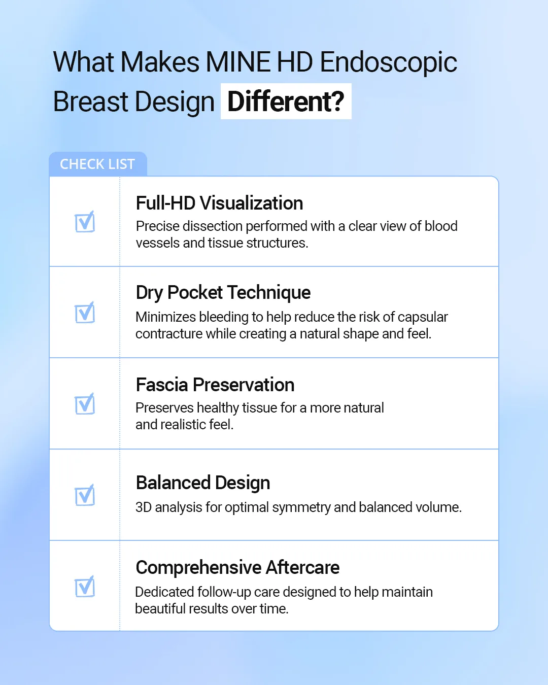
Dr. Lee’s key point: Of all the endoscopic breast augmentation benefits, the one that matters most is a more predictable result. It is a tool for precision, not a promise that nothing can go wrong.
Limits of Endoscopic Breast Augmentation: What a Camera Cannot Fix
Knowing the benefits of endoscopic breast augmentation is only half the picture. The other half is knowing where they stop.
It does not choose the implant or the plane for you
The camera improves what the surgeon can see. It does not decide the final shape. That still depends on implant size and shape, the pocket plane and the thickness of your own tissue. Textbooks explain that the choice between placing the implant above or below the muscle depends on the implant type and how much tissue covers it. A poorly planned operation with a good camera is still a poorly planned operation.
Not every patient or revision is suited to it
Suitability depends on your chest anatomy, skin and tissue thickness, and any previous surgery. In some revision cases, an incision under or around the breast gives better access to the problem than the armpit. This can only be decided after an in-person examination.
Side effects that still apply
Endoscopic surgery is still surgery. These side effects remain possible:
- Swelling and bruising: the most common part of recovery. Most swelling and bruising improves within 2–3 weeks.
- Hematoma: can occur within 1–2 weeks after surgery and needs prompt treatment. According to plastic surgery textbooks, a hematoma is treated by reoperation to drain the blood, stop the bleeding and wash the pocket, and the implant usually does not need replacing. Textbooks describe four warning signs: a breast that is swollen, painful, bruised and very tender to the touch.
- Infection or inflammation: uncommon, but in some cases the implant must be removed.
- Capsular contracture: firmness that can develop over time.
- Change in nipple sensation: temporarily reduced sensation is common and, in most cases at Mine, recovers within about six months.
- Implant malposition or rotation.
Dr. Lee’s key point: A clinic that tells you there are no side effects is less trustworthy than one that explains them clearly and has a plan for each.
Recovery Timeline and Preparation
Many patients tell me they had less swelling and discomfort than they expected, and in our experience recovery after endoscopic surgery tends to feel easier. Still, I do not promise a specific number of days saved, because every recovery is different.
Week-by-week recovery table
The table below shows the typical course at our clinic. Individual recovery varies.
| Time after surgery | What to expect | Daily activity |
|---|---|---|
| Day 0–3 | Swelling and discomfort at their peak | Rest is essential |
| Day 4–7 | Swelling begins to ease, pain decreases | Light daily activities; light office work from about day 5–7 |
| Weeks 1–2 | Bruising fades, stitches removed | Light office work; physically demanding jobs still need rest |
| Weeks 3–4 | Most swelling has settled | Most daily activities |
| Months 1–3 | Implant gradually settles into position | Light exercise |
| Months 3–6 | Final, natural shape develops | All activities |
Office workers can usually return to light work 5–7 days after surgery, while physically demanding jobs need 2–3 weeks or more. The final shape settles over 3–6 months.
Two weeks before surgery: what to stop
Plastic surgery textbooks recommend stopping medications and supplements that increase bleeding for at least 2 weeks before breast augmentation. At Mine, we also ask patients to prepare in these ways:
- Stop aspirin and supplements such as omega-3 and vitamin E, which can increase bleeding.
- Stop smoking and drinking alcohol at least two weeks before surgery, as both affect circulation and healing.
- Avoid scheduling surgery during your period if possible, as swelling can be worse.
- Complete the pre-operative blood tests and ECG (heart tracing).
- Tell us about every medication you take regularly.
After surgery, a few habits make a real difference:
- Wear the compression bra for as long as instructed. It helps the implant settle in place.
- Sleep with your upper body raised about 30 degrees to reduce swelling.
- Massage only when your medical team tells you to. Incorrect massage can contribute to implant rotation.
- Avoid strenuous exercise for at least one month.
- Wait about two months before using a sauna.
Frequently Asked Questions
Is endoscopic breast augmentation safer than standard breast augmentation?
It reduces specific risks linked to blind dissection, mainly bleeding and hematoma, because bleeding is controlled under direct vision. Textbooks support this advantage. It does not remove every surgical risk, and infection, capsular contracture and malposition remain possible.
How is an HD endoscope different from a standard endoscope?
Comparing our system with a standard endoscope, the HD endoscope offers higher resolution, a wider viewing angle and a brighter cold light source, which make small vessels and the pocket surface easier to see. This is our equipment explanation, not the finding of a published study.
Does endoscopic breast augmentation leave a scar?
Yes, a small scar in the armpit, but none on the breast itself. No surgery is completely scar-free. The armpit scar is placed in a natural crease where it is rarely noticed in daily life.
How long is recovery after endoscopic breast augmentation?
Most office workers return to light work 5–7 days after surgery, and physically demanding jobs need 2–3 weeks or more. Swelling largely settles in 3–4 weeks, and the final shape develops over 3–6 months.
Can endoscopic breast augmentation be used for revision surgery?
Often, yes, for problems such as capsular contracture or implant malposition, because the camera shows the old capsule and pocket. Depending on the problem, however, an incision on the breast may still be needed. An in-person examination is required to decide.
Does endoscopic breast augmentation affect breastfeeding?
Because the armpit approach does not cut directly through the milk-producing glandular tissue, it is generally considered to have little effect on breastfeeding. Individual differences exist, so please share any pregnancy plans during your consultation. For more detail, see our guide on whether breast surgery affects breastfeeding.
Planning Your Consultation
Endoscopic breast augmentation benefits are real, but they are specific: better bleeding control, a more precisely shaped pocket and an incision kept off the breast. What matters most is still a plan built around your chest structure, skin elasticity, tissue thickness and lifestyle. Even with the same implant and incision, the right pocket position and depth differ from person to person.
If you would like to know whether this approach suits you, you can read more about how Mine performs HD endoscopic breast augmentation, send your questions through our online consultation form, or call us at +82-2-516-1175. International patients can start with a preliminary review of photos, followed by an in-person consultation in Seoul.
Written and medically reviewed by Dr. Lee Sung-wook, Board-certified Plastic Surgeon (Korea), Mine Plastic Surgery, Seoul. Published October 2, 2026. Last reviewed October 2, 2026.
※ This content is for informational purposes only and does not replace professional medical advice, diagnosis, or treatment. Individual results may vary, and side effects may occur. Please consult with a board-certified plastic surgeon for personalized advice.