EN
EN


Acne scar types fall into four clinical groups: post-inflammatory hyperpigmentation (PIH — flat marks where only the color changed), atrophic (indented) scars, hypertrophic (raised) scars, and keloids (raised scars that spread past the original spot). What separates them is one thing — whether the skin lost collagen while healing or made too much of it.
If you are standing at the mirror trying to work out which marks will fade on their own and which ones are staying, that single distinction is the answer. A color change settles. Structural damage does not.
This guide does two things. First, it helps you identify which type of acne scar you are actually looking at. Second, it shows what that specific type needs. Acne scars are what inflammation leaves behind once the breakout itself has calmed, so if you are still dealing with active spots, start with how we treat active body acne first.
Table of Contents
- What Are Acne Scar Types? The Collagen Rule That Sorts All Four
- Acne Marks vs Acne Scars: The Touch Test That Tells Them Apart
- The Three Atrophic Types: Ice Pick, Boxcar and Rolling
- Raised Acne Scars: Hypertrophic vs Keloid
- Why Acne Scars on the Back and Chest Behave Differently
- Acne Scar Types and Treatment: What Each Type Actually Needs
- Risks, Skin Tone and What Results Are Realistic
- Frequently Asked Questions About Acne Scar Types
- Talk to a Board-Certified Specialist in Seoul
What Are Acne Scar Types? The Collagen Rule That Sorts All Four
Dermatology textbooks do not sort acne scars by how they look. They sort them by what happened to the collagen underneath. Collagen is the scaffolding protein that gives skin its thickness and its bounce, and it lives in the dermis — the living layer sitting below the surface you can see. Scars are decided down there, not on top.
Collagen lost vs collagen overmade
When inflammation from a breakout damages the dermis, healing goes one of two ways. If the skin ends up with less collagen than it started with, the surface has nothing holding it up and it sinks. That is an atrophic scar — a dent. If the repair process overshoots and lays down more collagen than the wound needed, the tissue rises above the surrounding skin. That is a hypertrophic scar or, in its more aggressive form, a keloid.
So the four groups this article uses are: PIH, atrophic, hypertrophic and keloid. One of those four is the odd one out. Post-inflammatory hyperpigmentation is not structurally a scar at all — the skin architecture is intact and only the pigment has changed. It gets grouped in because patients see it as a scar, and because treating it as one is a common and costly mistake.
Why most acne scarring is the indented kind
Roughly 80 to 90 percent of people with acne scarring have atrophic scars, the indented kind, while hypertrophic scars and keloids account for the minority. That imbalance is why so much acne scar treatment is built around rebuilding lost volume rather than reducing excess tissue.
Classification is not academic housekeeping. As the American Academy of Dermatology notes, the approach that improves one type can do nothing for another — and in some cases makes it worse. Telling these four types apart is the first clinical step, not an optional extra.
Acne Marks vs Acne Scars: The Touch Test That Tells Them Apart
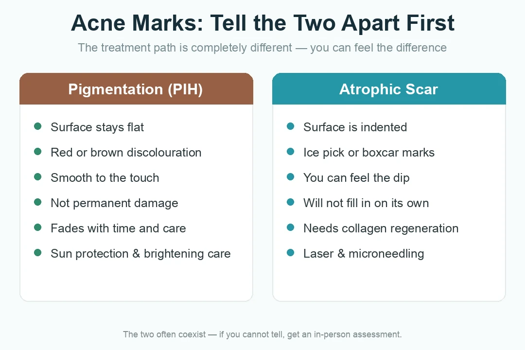
Here is the test that costs nothing and settles most of the confusion. Close your eyes and run a fingertip slowly across the mark. If the surface feels smooth and even and only the color is different, you are touching pigment. If you can feel the skin dip away under your finger, you are touching a structural scar.
Post-inflammatory hyperpigmentation (PIH): flat, discolored, temporary
PIH is a pigment change. The spot inflamed the skin, the skin responded by producing extra melanin in that patch, and the color stayed behind after the spot itself resolved. The surface stays flat. The color reads as brown or slate-gray, and it is more pronounced and more persistent in deeper skin tones.
Some post-acne marks are flat and red rather than brown. That redness comes from blood vessels left dilated in the healed skin, and it behaves differently from melanin-driven marks. Either way, the defining feature is the same: the surface is smooth, the damage is not structural, and the mark fades over time with consistent sun protection.
Atrophic scars: indented, and you can feel the dip
An atrophic scar is a genuine loss of tissue. The floor of the mark sits lower than the skin around it because the collagen that used to hold it up is gone. Time alone does not fix this. The dent will still be there in five years unless collagen is deliberately rebuilt in that spot.
In practice the two coexist constantly. The same patch of cheek or shoulder often carries flat pigment on top of genuinely indented tissue, which is exactly why self-diagnosis is unreliable. It is also why the two most expensive mistakes in this field happen: aggressive resurfacing aimed at what is really pigment, and brightening creams applied faithfully for months to what is really a dent. An in-person assessment under proper lighting sorts this out in minutes.
The Three Atrophic Types: Ice Pick, Boxcar and Rolling
Of the four acne scar types, the atrophic group is the only one that subdivides further. Atrophic acne scars come in three shapes, and the shape decides what can reach them. This three-way division comes from a peer-reviewed classification system for acne scars published in 2001, which sorted them by width, depth and three-dimensional architecture specifically so that scar anatomy could be matched to treatment that actually reaches it.
Ice pick scars — narrow, deep, under 2 mm
Ice pick scars make up 60 to 70 percent of atrophic acne scars, measure under 2 millimeters across, and cut a narrow V-shaped tract down into the dermis. In some cases the tract reaches the fatty tissue below it.
Picture something narrow pushed straight down into the skin: a small, sharply outlined opening at the surface with a channel running deep beneath it. That geometry is the whole problem. The opening is tiny, so the visible mark looks minor, but the damage extends far deeper than any surface-level treatment can reach.
Boxcar scars — round or oval, steep-walled, 1.5–4 mm
Boxcar scars account for 20 to 30 percent of atrophic acne scars, run 1.5 to 4 millimeters across, and are graded as shallow when they sit under 0.5 millimeters deep and deep when they exceed it.
Think of a shallow crater with steep sides and a flat, wide floor. Unlike ice pick scars, boxcar scars do not taper to a point at the bottom — the base stays broad. They are also visibly wider at the surface. Shallow boxcar scars respond far more readily than deep ones, which is why depth grading changes the plan even within a single scar type.
Rolling scars — wide, soft-edged, over 4 mm
Rolling scars are the widest of the three at more than 4 to 5 millimeters, and form when otherwise normal skin is tethered downward by fibrous bands from underneath. They make up roughly 15 to 25 percent of atrophic scars.
This one is counterintuitive. The skin at the surface is essentially healthy — it is being pulled down from below by abnormal fibrous anchors, which creates a soft, undulating, wave-like shadow rather than a sharp-edged pit. That is also why rolling scars seem to appear and disappear depending on where you stand. Under flat, direct light they can be almost invisible. Under angled or indirect light, the shadows they cast make them obvious.
One point of clinical honesty matters more than any of the measurements above: all three subtypes can be deep, and they routinely coexist in the same person on the same area of skin. A single clean diagnosis is genuinely difficult even for clinicians examining skin directly. Reading a list like this one will orient you, but it will not replace an examination.
Raised Acne Scars: Hypertrophic vs Keloid
The two raised acne scar types sit on the collagen-gain side of the rule from earlier. The body did not under-repair the wound — it over-repaired it, laying down dense fibrous tissue that pushes the surface upward instead of letting it sink.
Hypertrophic: raised, but it stays inside the original border
A hypertrophic scar is thickened and raised, but it respects the boundary of the original blemish. Trace the outline of where the spot used to be, and the raised tissue stays inside it. These scars often soften and flatten somewhat over a year or two, though they rarely settle completely on their own.
Keloid: it grows past where the spot ever was
A keloid does not respect that boundary. The overgrown tissue spreads outward into skin that was never inflamed, so a keloid ends up larger than the spot that started it. That single question — does it stay inside the original footprint or push beyond it? — is what separates the two clinically.
Skin tone matters here in a way it does not for atrophic scars. Hypertrophic scars and keloids are reported in deeper skin tones at rates 3 to 18 times higher than in lighter ones, which is why an honest treatment plan starts with your skin type rather than with a machine. Raised scars are also less common on the face than indented ones, but the chest, shoulders and back are classic keloid-prone territory.
Raised and keloid scars follow a different pathway from indented ones — one closer to scar revision than to skin resurfacing. If that is what you are dealing with, read how raised and keloid scars are treated and our page on surgical scar revision.
Why Acne Scars on the Back and Chest Behave Differently
Thicker skin, more oil, constant tension
Patients often ask why the marks on their back and shoulders look worse, last longer and scar more readily than anything on their face. The answer is anatomy, and it is fairly straightforward.
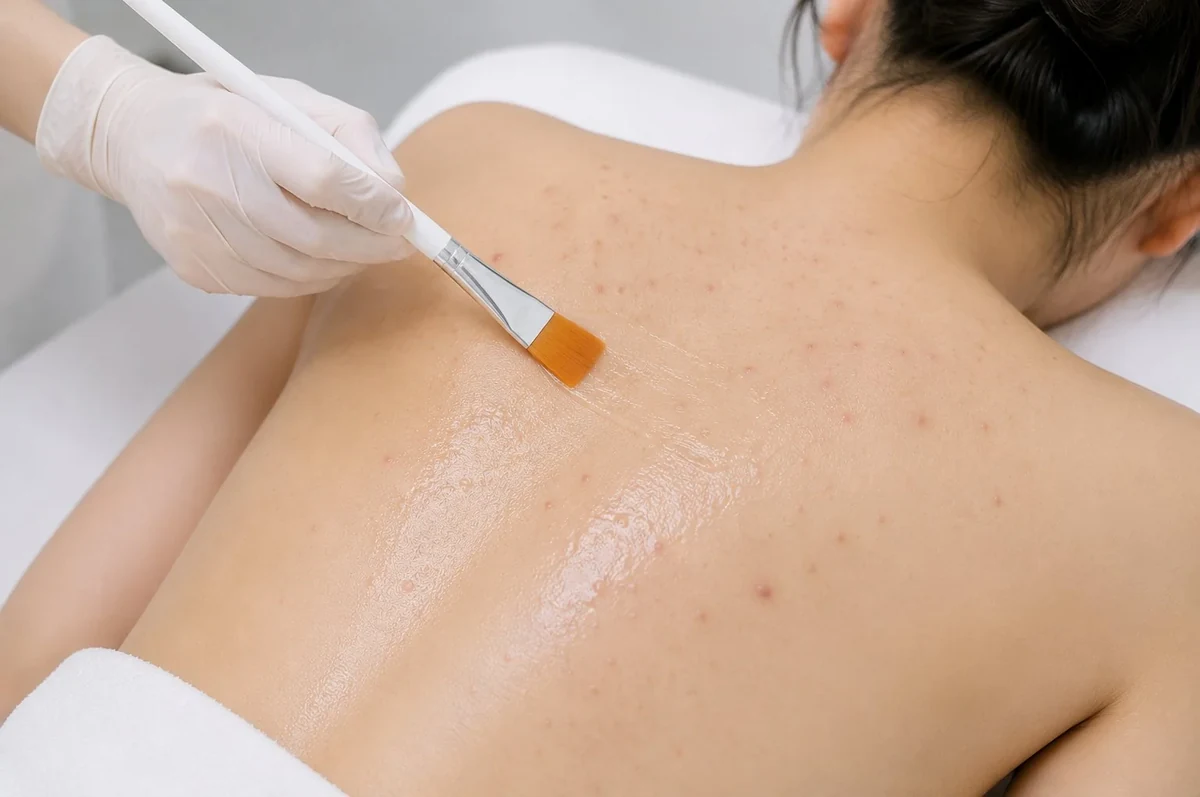
Back skin carries the highest density of oil glands on the body, and it has both a thicker outer layer and a thicker dermis than facial skin. Inflammation there has further to travel and more fuel to work with, so it tends to be driven deeper before it resolves. Deeper inflammation means more dermal involvement, and dermal involvement is what turns a spot into a scar.
Then there is tension. The back and shoulders are under sustained mechanical load from ordinary movement — every reach, twist and stretch pulls on healing skin. Mechanical tension is the classic driver of keloid formation, which is why raised acne scars skew disproportionately toward the trunk. Friction from clothing, backpack straps and sports gear adds a second insult, re-irritating skin that is trying to close.
Timing compounds all of it. The outer layer of skin renews in about 28 days, but once the dermis is involved a scar needs 6 to 12 months to finish maturing — which is why marks on the back outlast the ones on your face.
One clarification before moving on: if what you have is still active acne on the back or chest rather than its aftermath, that is a different treatment path entirely, and medical-grade in-clinic peels are the option we use for it. That territory is covered in full in our guide to treating active body acne.
Acne Scar Types and Treatment: What Each Type Actually Needs
This is where identification pays off. Each of the types described above needs something structurally different, and the mismatches are not neutral — several of them actively set you back.
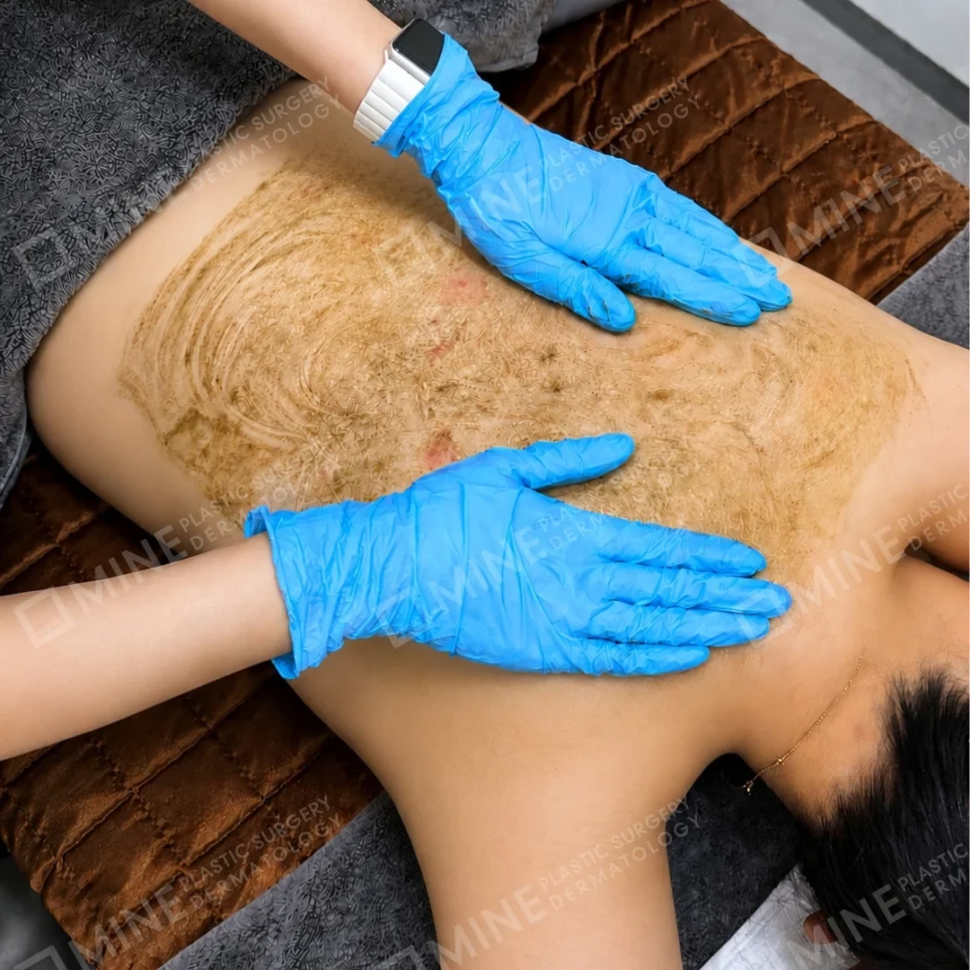
Flat pigment (PIH): protect, fade, do not resurface
For flat pigment marks, sun protection is not a supporting measure. It is the treatment. Ultraviolet exposure re-darkens the pigment faster than any topical product fades it, so daily protection is what decides whether the mark clears in months or persists for years. Brightening care and gentle turnover support help from there.
What does not help is aggressive resurfacing. Treating active pigment with a device that creates controlled injury can trigger more pigment production, particularly in deeper skin tones. This is the single most common way patients make marks worse while trying to improve them.
Ice pick and deep boxcar: focused, depth-reaching approaches
These scars fail surface-level treatment for a mechanical reason: the opening is narrow and the damage is deep, so anything working on the top layers never reaches the bottom of the tract. Ablative fractional laser resurfacing, which creates controlled columns of injury that penetrate to a set depth and trigger collagen rebuilding as they heal, is the textbook mainstay for this reason. Deep boxcar scars generally need repeated, staged treatment rather than a single session.
Rolling: release the tether first, then rebuild collagen
Rolling scars have a distinct sequence. Because the surface is normal skin being pulled from underneath, resurfacing it while the fibrous anchor is still attached achieves very little — the skin is released briefly and then pulled straight back down. The tether has to be released first. Only then does collagen stimulation make sense, whether through microneedling or through skin regeneration options for indented scars such as polynucleotide treatment, which uses purified DNA fragments to support the skin’s own repair activity.
Raised and keloid: flatten, do not resurface
Raised scars invert the goal completely. Here the aim is to reduce and flatten overgrown tissue rather than stimulate new collagen — which is precisely why applying an indented-scar protocol to a raised scar can enlarge it. Skin tone raises the stakes further, given the higher keloid tendency noted earlier.
| Scar type | What you see | What you feel | What it needs | Realistic course |
|---|---|---|---|---|
| PIH (flat pigment) | Brown, gray or red flat patch | Smooth — no dip | Sun protection, brightening and gentle turnover care | Gradual fading over months; no resurfacing |
| Ice pick | Small pinpoint opening, under 2 mm | Sharp, narrow pit | Depth-reaching focused treatment | Staged sessions; improvement, not removal |
| Boxcar | Round or oval crater, 1.5–4 mm | Steep walls, flat floor | Resurfacing matched to depth grading | Shallow responds faster than deep |
| Rolling | Wide wavy shadow, over 4 mm | Soft undulation, no sharp edge | Release the tether, then rebuild collagen | Sequenced over several months |
| Hypertrophic | Raised, within the original border | Firm, thickened ridge | Flattening and softening approaches | Slow; may partly settle on its own |
| Keloid | Raised, spreading past the original spot | Firm, sometimes itchy or tender | Scar-revision pathway, not resurfacing | Long-term management, recurrence possible |
One thing the table cannot show is how often these overlap. Because the three atrophic subtypes routinely coexist, real treatment plans are almost always combinations staged across months rather than a single device applied repeatedly. Matching acne scar types to treatment is a clinical assessment, not something a mirror and a checklist can finish — which is what acne and acne scar care at MINE is built around.
Risks, Skin Tone and What Results Are Realistic
Side effects you should expect to be told about
Every approach described above carries risks, and a consultation that does not name them is incomplete. Resurfacing procedures commonly cause redness and swelling for several days, along with crusting and peeling as the skin repairs. Temporary darkening of the treated area is common. Infection is possible whenever the skin barrier is broken, and pigment change — either darker or lighter than surrounding skin — is a real risk rather than a theoretical one.

Why deeper skin tones need a more cautious plan
Patients with deeper skin tones carry a higher risk of post-inflammatory hyperpigmentation caused by the treatment itself, alongside the higher keloid tendency mentioned earlier. Device settings, energy levels and the spacing between sessions all have to be adjusted accordingly. It is also worth stating plainly that comparative research in skin of color remains limited, and no single universal protocol has been established — which is an argument for conservative, staged treatment under experienced supervision.
On results: meaningful reduction over a course of treatment is a reasonable expectation. A return to skin that never scarred is not. Individual results vary, and honest planning starts from that. Our discussion of what realistic scar improvement looks like sets out the same framing in more detail. All treatment plans at MINE Plastic Surgery & Dermatology in Seoul are reviewed by Dr. Lee Sung-wook, a board-certified plastic surgeon.
Frequently Asked Questions About Acne Scar Types
How do I know if I have acne scars or just acne marks?
Use the touch test. Run a fingertip over the mark with your eyes closed. If the surface feels smooth and only the color is different, it is post-inflammatory hyperpigmentation, which fades over time with sun protection. If you can feel the skin dip beneath your finger, it is an atrophic scar, and it will not fill in on its own no matter how long you wait. Bear in mind that the two frequently sit side by side on the same area, which is why a clinical assessment is more reliable than a mirror.
How long do acne marks take to fade on their own?
Flat pigment marks fade gradually over months as the skin turns over. The outer layer renews in roughly 28 days, but pigment sits deeper than that, so the visible change is slower than the turnover cycle suggests. Daily sun protection is the single biggest variable, because ultraviolet exposure re-darkens the pigment faster than it fades. Structural scars are a different matter entirely — an indented scar does not improve with time, because the collagen loss underneath it is permanent unless it is deliberately rebuilt.
Why are acne scars on my back and chest worse than on my face?
Back skin has the highest oil-gland density on the body plus a thicker dermis, so inflammation is driven deeper before it settles, and deeper inflammation means more scarring. The back and shoulders are also under constant mechanical tension from ordinary movement, along with friction from clothing and bags. Tension is the classic driver of raised and keloid scarring, which is why acne scars on the back and chest skew toward the raised types while facial ones skew indented.
How many types of atrophic acne scars are there?
Three, in standard dermatology classification. Ice pick scars account for 60 to 70 percent, measure under 2 millimeters across and run narrow and deep. Boxcar scars account for 20 to 30 percent, measure 1.5 to 4 millimeters, and have steep walls with a flat floor. Rolling scars account for 15 to 25 percent, exceed 4 to 5 millimeters, and are tethered downward from underneath. Most patients have a mixture rather than one clean type, which is why treatment plans are usually combinations.
Can one treatment fix every acne scar type at once?
No — and that is the core reason scar type matters. Indented scars need collagen rebuilt, raised scars need overgrown collagen reduced, and flat pigment needs neither. Applying the wrong approach can make a raised scar or a pigment mark worse rather than leaving it unchanged. Realistic plans stage several approaches across months, and they begin with an in-person assessment rather than with a device selection.
Talk to a Board-Certified Specialist in Seoul
If you take one thing from this guide, take this: work out which type of acne scar you have first. The approach that lifts a dent will do nothing for a pigment mark, and the approach that fades a pigment mark will do nothing for a dent.
That identification is a clinical step. It needs proper lighting, direct examination and someone who looks at post-acne skin daily. International patients can begin with a photo-based pre-assessment before traveling, and we will tell you honestly what a course of treatment can and cannot change. You can start with acne and acne scar care at MINE.
※ This content is for informational purposes only and does not replace professional medical advice, diagnosis, or treatment. Individual results may vary, and side effects may occur. Please consult with a board-certified plastic surgeon for personalized advice.