EN


“I had salivary gland Botox and nothing changed.”
“I lost weight, but the fullness under my jaw stayed.”
“My face looks wider in photos than it does in the mirror.”
These sound like three different problems, but in my clinic they often share one cause: an enlarged salivary gland that was never clearly identified, or a toxin that never reached it.
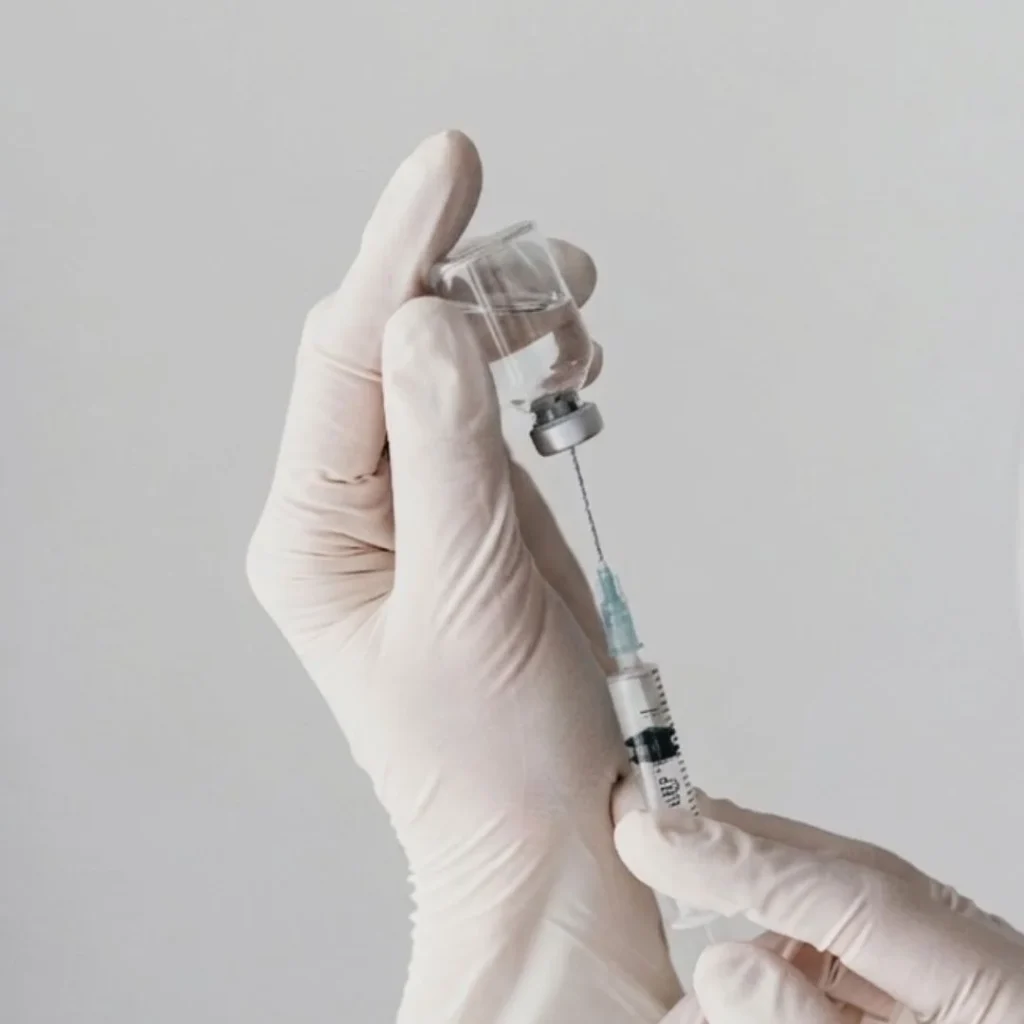
Ultrasound-guided salivary gland Botox is an injection of botulinum toxin into an enlarged salivary gland while the doctor watches the gland, its depth and the nearby structures on a live ultrasound screen, so the toxin goes inside the gland rather than beside it. The toxin itself is the same one used in a blind injection. What changes is where it lands.
In this guide, I explain how ultrasound-guided salivary gland Botox differs from an injection placed by feel, why a treatment sometimes “doesn’t work,” and how we decide whether the gland or the jaw muscle should be treated. Ultrasound is a helpful tool, but an accurate diagnosis is the real first step.
Why an Enlarged Salivary Gland Changes Your Jawline
Where the submandibular and parotid glands sit
The submandibular gland sits just under the inner edge of the jawbone, right in the area most people call the double chin. Its job is to make saliva and send it into the mouth. Its size varies a lot from person to person.
When this gland is large, the area under the jaw looks full and rounded. It is easy to mistake for fat, but the volume comes from gland tissue. That explains why some people notice:
- a soft bulge under the jaw that does not go away with weight loss
- a blurred border between the jaw and the neck
- a heavy-looking jawline in side-view photos
- little change after fat removal alone
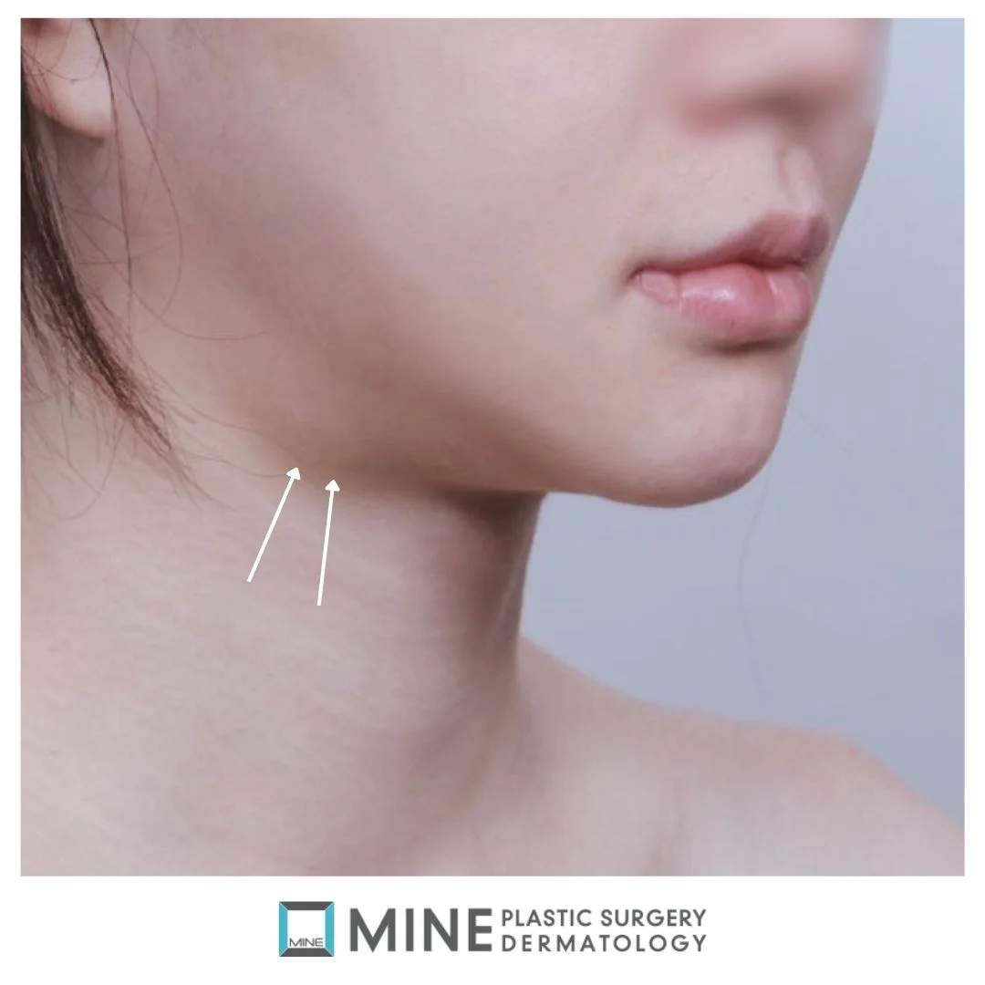
The parotid gland sits in front of and below the ear, and in some people it partly wraps around the masseter (the main chewing muscle). Treating the parotid is a different plan from treating the gland under the jaw; our guide to parotid vs submandibular treatment covers that choice.
Does chewing make salivary glands bigger?
This is one of the most common questions I hear. Chewing mainly works the masseter (the jaw muscle), not the salivary gland, so chewing habits are rarely the main reason a gland is large. In clinical practice, an enlarged submandibular gland usually reflects a mix of factors: an inherited larger gland, a nervous system that tends to drive more saliva, and long-term irritation or inflammation. Because of this, changing your diet or massaging the area will not shrink the gland itself.
Gland, masseter, fat or platysma: four look-alikes
Fullness under and along the jaw can come from four different structures: the salivary gland, the fat under the skin, the platysma (the thin sheet of muscle in the neck), or a large masseter and jaw shape. From the outside, they can look almost the same. Each one needs a different treatment, which is why the target has to be confirmed before any toxin is used.
Blind vs Ultrasound-Guided Salivary Gland Botox: What Actually Differs
Before comparing the two methods, it helps to know how the toxin works. Botulinum toxin blocks acetylcholine, the nerve signal that tells a salivary gland to make saliva. With less of that signal, the gland becomes less active and gradually gets smaller. This mechanism is well described in medical textbooks and salivary gland research.
For that effect to happen, the toxin has to end up in the gland. Here is how the two approaches compare:
| Blind (palpation / landmark) injection | Ultrasound-guided salivary gland Botox | |
|---|---|---|
| Locating the gland | By feel and surface landmarks | Gland outline, depth and size seen on screen |
| Depth control | Estimated | Needle tip and gland depth visible |
| Nearby nerves and vessels | Avoided based on experience | Branches near the gland (such as the marginal mandibular branch of the facial nerve) and vessels identified before injecting |
| Dose | Often a standard amount | Planned from the measured gland size |
| Repeatability | Depends on memory and notes | The same points can be found again next time |
What a blind (palpation-based) injection relies on
A blind injection relies on the injector’s hands and on surface landmarks such as the angle of the jaw. Experienced injectors can do this well. Still, the submandibular gland sits deep and is wrapped in a layer of connective tissue (fascia), so the depth of the needle tip is always partly an estimate.
What real-time ultrasound adds
With ultrasound guidance, the gland shows up on the screen as a distinct shape. We can see how deep it is, how big it is, and where the needle tip sits before the toxin goes in.
A salivary-gland surgery textbook describes splitting the dose across 4–10 small points in a parotid gland, depending on its size, preferably under ultrasound control. The same textbook gives 0.1–0.2 mL as the small amount placed at each point. That protocol was written for a different medical problem, not for cosmetic slimming, but it shows that placing toxin inside a gland in small, guided portions is the approach the literature prefers.
What ultrasound cannot do
Ultrasound does not make side effects impossible, and it does not replace diagnosis. If the fullness under your jaw is mainly fat or muscle, a perfectly placed injection into the gland will still disappoint. The scan supports good judgment; it does not stand in for it.
“It Didn’t Work”: When the Toxin Lands Outside the Gland
Some patients come to us after treatment elsewhere saying they saw no change. When we look more closely, the cause usually falls into one of two groups: the toxin did not stay in the gland, or the gland was not the right target in the first place.
A deep gland wrapped in fascia
Anatomy textbooks note that the submandibular gland sits deep inside a fascia layer, with the mylohyoid muscle (a muscle of the floor of the mouth) passing through it. If toxin spreads beyond the gland, it can affect the muscles used for swallowing. That is why injection depth matters so much here, and why we always guide these injections with ultrasound. You can read more in our article on submandibular gland Botox and swallowing.
When the parotid and masseter overlap
The parotid gland lies under a firm layer of fascia and has its own capsule, which can resist an injection. Because it can partly surround the masseter, toxin meant for the muscle can end up in the gland, and the reverse can also happen. Anatomy texts also describe the facial nerve running between the two lobes of the parotid, which is one more reason to see the gland clearly before injecting.
Wrong target vs wrong placement
In my experience, the deciding factor is often not whether ultrasound was used, but whether the diagnosis before treatment was right. We see two common patterns:
- The gland was treated even though fat, the platysma or the jaw structure was the main cause of the fullness.
- The gland really was enlarged, but the dose was too small for its size.
Ultrasound helps us check these structures more objectively. For a full checklist of other causes, see why salivary gland Botox sometimes doesn’t work.
Choosing the Injection Target: Salivary Gland or Masseter?
A large masseter and an enlarged salivary gland can look surprisingly similar from the outside. The two sit at different depths and in different places, so the injection points must be different too.
How the scan tells gland from muscle
During the exam, we often ask you to clench your teeth. The masseter hardens under the fingers, while the gland does not. Ultrasound then confirms what we feel: muscle and gland tissue look different on the screen, and we can measure each one. This post focuses on ultrasound for guiding the needle; if you want to know more about the scan itself, read how gland enlargement is diagnosed on ultrasound.
When both are treated in one visit
Many patients have both a strong masseter and a prominent gland. Both can be treated in the same visit, with separate injection points and a separate dose for each structure.
Medical literature also notes that a prominent parotid gland can become more noticeable after masseter Botox slims the jaw muscle. This is another reason we look at the whole lower face, not just one structure, before planning treatment.
How MINE Performs Ultrasound-Guided Salivary Gland Botox, Step by Step
At MINE, the guided technique follows 4 steps: scan, map nearby structures, plan the dose, and inject under live ultrasound.
- Consultation and scan. Dr. Lee examines your face and neck and scans the gland to check its position, depth and size.
- Mapping nearby structures. Nerves and blood vessels around the gland are identified on the screen, including nearby nerve branches such as the marginal mandibular branch of the facial nerve, and a safety margin is set.
- Planning the dose. The dose is set for your gland’s size and how much it sticks out. This avoids putting too much into a small gland or too little into a large one.
- Injecting under live ultrasound. The toxin is divided across several points, and we confirm on the screen that it enters the gland.
A numbing cream can be applied beforehand if you prefer. Because the measurements are recorded, the same points can be located again at your next session.
Recovery, Side Effects and Results at a Glance
These topics each have their own detailed guide, so here is a short summary:
- Side effects: Most are mild and short-lived, such as swelling, tenderness or bruising at the injection points, and some people notice a drier mouth for a while. Rarely, weakness related to a nearby nerve can occur; if your smile or lower lip feels different, contact us promptly. Read more about the effects and side effects of salivary gland Botox.
- Aftercare: Simple aftercare steps and a few habits to pause after treatment help the toxin settle where it was placed.
- Results and duration: Changes appear gradually; see when results start to show, how long the effect lasts and the signs it is wearing off. The effect is temporary, so repeat treatment is part of the plan.
- Cost: Our guide explains what affects the cost.
- Tell us first if: you are pregnant or breastfeeding, have a neuromuscular or autoimmune condition, take blood thinners, or have had gland or masseter Botox elsewhere.
Frequently Asked Questions
Is ultrasound-guided salivary gland Botox more effective than a blind injection?
It improves the chance that the toxin is placed inside the gland at the right depth, which is what the treatment depends on. It is not a guarantee, though. The result still depends on a correct diagnosis and an appropriate dose for your gland’s size.
Why didn’t my salivary gland Botox work?
The most common reasons are that the gland was not the main cause of the fullness, the dose was too low for the gland’s size, or the toxin ended up outside the gland. Our article on salivary gland Botox not working goes through each cause.
How do doctors tell an enlarged salivary gland from a big masseter?
The two sit in different places and at different depths. A clench test shows the muscle hardening, and ultrasound shows gland tissue and muscle as clearly different structures that can each be measured.
Can masseter Botox and salivary gland Botox be done together?
Yes. Both can be treated in one visit, using separate injection points and a separate dose for each. Ultrasound helps keep the two targets apart.
Does ultrasound-guided salivary gland Botox hurt or need downtime?
It is a needle injection, and numbing cream can be used if you are sensitive. Most people return to normal activities the same day, though saunas, intense exercise and alcohol should be avoided for a short time. Our aftercare guide has the details.
Book an Ultrasound Assessment at MINE
Accurate treatment starts with an accurate diagnosis. Before any injection, we confirm whether the gland, the masseter, fat or the platysma is behind the fullness, and only then decide whether ultrasound-guided salivary gland Botox is the right option for you.
At MINE Plastic Surgery in Gangnam, Seoul, board-certified plastic surgeon Dr. Lee Sung-wook personally performs the scan and plans each injection. Learn more about salivary gland reduction at MINE, or use the consultation form on this website or call +82-2-516-1175 to book an assessment.
Written and reviewed by Dr. Lee Sung-wook, board-certified plastic surgeon, MINE Plastic Surgery.
※ This content is for informational purposes only and does not replace professional medical advice, diagnosis, or treatment. Individual results may vary, and side effects may occur. Please consult with a board-certified plastic surgeon for personalized advice.